Authors:Fang Zige, Yang Zefeng, Liu Xiaoyi, Xie Bin, Peng Yuying, Liu Yuhong, Liang Xiaohong, Lin Fengbin, Song Yunhe, Zhang Xiulan
Corresponding author:Zhang Xiulan,Email:zhangxl2@mail.sysu.edu.cn
Published:2026-08-10
DOI:10.3760/cma.j.cn115989-20260212-00077
ABSTRACT
Objective To evaluate alterations in macular microvascular fractal dimension (FD) and blood vessel tortuosity (BVT) in patients with primary open-angle glaucoma (POAG) using optical coherence tomography angiography (OCTA).
Methods A cross-sectional study was conducted. Early POAG patients (101 cases, 101 eyes) and healthy subjects (518 cases, 518 eyes) who underwent OCTA at Zhongshan Ophthalmic Center between July 2016 and July 2023 were included. Macular microvascular geometric parameters of the superficial capillary plexus (SCP) and deep capillary plexus (DCP) were obtained from OCTA images and compared between two groups. Univariate and multivariate regression models were used to identify ocular factors associated with FD and BVT. This study followed the Declaration of Helsinki. The study protocol was approved by the Ethics Committee of Zhongshan Ophthalmic Center, Sun Yat-sen University (No. 2023 KYPJ356-4), and all subjects signed the informed consent form.
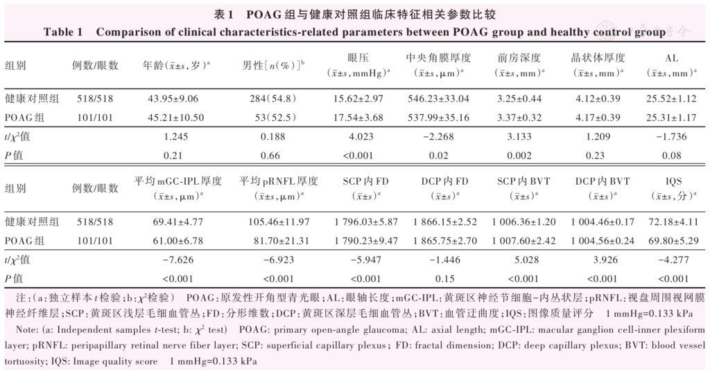
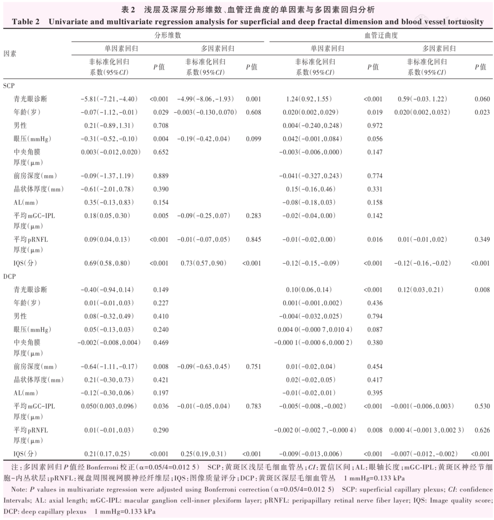
Results Compared with healthy control group, POAG group showed significantly lower FD in the SCP and higher BVT in both the SCP and DCP ( t=-5.947、5.028、3.926; all P<0.001). In the multivariate model, diagnosis of glaucoma and image quality score (IQS) were influencing factors for FD in the SCP (diagnosis of glaucoma: β=-4.99, 95% confidence interval[ CI]: -8.06–1.93, P=0.001; IQS: β=0.73, 95% CI: 0.57-0.90, P<0.001) and BVT in the DCP (diagnosis of glaucoma: β=0.12, 95% CI: 0.03-0.21, P=0.008; IQS: β=-0.007, 95% CI: -0.012–0.002, P<0.001).
Conclusions Compared with healthy controls, there were significant differences in FD and BVT of macular microvessels in patients with early POAG, and the diagnosis of POAG is associated with macular microvascular geometry parameters.
KEYWORDS:
Glaucoma;Optical coherence tomography angiography;Fractal dimension;Blood vessel tortuosity;Determinants
COPYRIGHTS:
Copyright by Chinese Medical Association
No content published by the journals of Chinese Medical Association may be reproduced or abridged without authorization. Please do not use or copy the layout and design of the journals without permission.
All articles published represent the opinions of the authors, and do not reflect the official policy of the Chinese Medical Association or the Editorial Board, unless this is clearly specified.
Authors Info & Affiliations
Fang Zige
State Key Laboratory of Ophthalmology, Zhongshan Ophthalmic Center, Sun Yat-sen University, Guangdong Provincial Key Laboratory of Ophthalmology and Visual Science, Guangdong Provincial Clinical Research Center for Ocular Diseases, Guangzhou 510060, China
Yang Zefeng
State Key Laboratory of Ophthalmology, Zhongshan Ophthalmic Center, Sun Yat-sen University, Guangdong Provincial Key Laboratory of Ophthalmology and Visual Science, Guangdong Provincial Clinical Research Center for Ocular Diseases, Guangzhou 510060, China
Liu Xiaoyi
State Key Laboratory of Ophthalmology, Zhongshan Ophthalmic Center, Sun Yat-sen University, Guangdong Provincial Key Laboratory of Ophthalmology and Visual Science, Guangdong Provincial Clinical Research Center for Ocular Diseases, Guangzhou 510060, China
Xie Bin
State Key Laboratory of Ophthalmology, Zhongshan Ophthalmic Center, Sun Yat-sen University, Guangdong Provincial Key Laboratory of Ophthalmology and Visual Science, Guangdong Provincial Clinical Research Center for Ocular Diseases, Guangzhou 510060, China
Peng Yuying
State Key Laboratory of Ophthalmology, Zhongshan Ophthalmic Center, Sun Yat-sen University, Guangdong Provincial Key Laboratory of Ophthalmology and Visual Science, Guangdong Provincial Clinical Research Center for Ocular Diseases, Guangzhou 510060, China
Liu Yuhong
State Key Laboratory of Ophthalmology, Zhongshan Ophthalmic Center, Sun Yat-sen University, Guangdong Provincial Key Laboratory of Ophthalmology and Visual Science, Guangdong Provincial Clinical Research Center for Ocular Diseases, Guangzhou 510060, China
Liang Xiaohong
State Key Laboratory of Ophthalmology, Zhongshan Ophthalmic Center, Sun Yat-sen University, Guangdong Provincial Key Laboratory of Ophthalmology and Visual Science, Guangdong Provincial Clinical Research Center for Ocular Diseases, Guangzhou 510060, China
Lin Fengbin
State Key Laboratory of Ophthalmology, Zhongshan Ophthalmic Center, Sun Yat-sen University, Guangdong Provincial Key Laboratory of Ophthalmology and Visual Science, Guangdong Provincial Clinical Research Center for Ocular Diseases, Guangzhou 510060, China
Song Yunhe
State Key Laboratory of Ophthalmology, Zhongshan Ophthalmic Center, Sun Yat-sen University, Guangdong Provincial Key Laboratory of Ophthalmology and Visual Science, Guangdong Provincial Clinical Research Center for Ocular Diseases, Guangzhou 510060, China
Zhang Xiulan
State Key Laboratory of Ophthalmology, Zhongshan Ophthalmic Center, Sun Yat-sen University, Guangdong Provincial Key Laboratory of Ophthalmology and Visual Science, Guangdong Provincial Clinical Research Center for Ocular Diseases, Guangzhou 510060, China
Figures & Tables
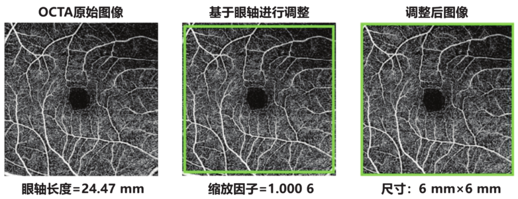
Figure 1 Magnification correction by using Littmann and Bennett formula Scaling factor=3.382×0.013 062×(AL-1.82). The scaling factor was 1 if AL= 23.82 mm. En-face image of superficial capillary plexus at macula with AL=24.47 mm was presented and the scaling factor was calculated as 1.00 006. The green square corresponded to the actual image to the size of 6 mm×6 mm. The cropped images were involved in the final analysis OCTA: optical coherence tomography angiography; AL: axial length
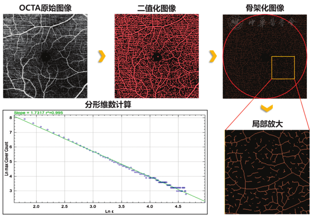
Figure 2 Schematic illustration of the retinal microvascular fractal dimension measurement and parameter extraction OCTA images and the macula were automatically determined in B-scan images. Scan size of 6 mm×6 mm of an en-face blood flow image of both superficial and deep retinas were stratified. Original images with same scan size following projection artifacts removal were obtained. Then binarized and skeletonized capillary images were calculated. In the log-log plot, the blue dot represented actual OCTA data for this image, while the green line displayed the closest fitting fractal log-log line. The x-axis indicated the log base e of the size of the boxes in pixels, while the y-axis represented the log base e of the number boxes subtending the OCTA pattern. Thus, a linear relationship in the log-log plot indicating the good self-similarity of the macular retinal microvascular network OCTA: optical coherence tomography angiography
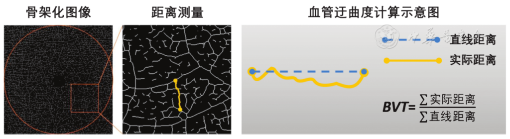
Figure 3 Schematic illustration of the retinal microvascular blood vessel tortuosity measurement and parameter extraction After binarization and skeletonization of the capillary OCTA images, distance parameters for calculating blood vessel tortuosity were extracted. Blood vessel tortuosity was calculated as the sum of the actual path lengths of the vessel branches divided by the sum of their straight-line distances BVT: blood vessel tortuosity; OCTA: optical coherence tomography angiography
References click to collapse
[1] Jayaram H, Kolko M, Friedman DS, et al. Glaucoma: now and beyond[J]. Lancet, 2023, 402(10414):1788‑1801. DOI: 10.1016/S0140‑6736(23)01289‑8.
[2] Taylor HR. Global blindness: the progress we are making and still need to make[J]. Asia Pac J Ophthalmol (Phila), 2019, 8(6):424‑428. DOI: 10.1097/APO.0000000000000264.
[3] Moghimi S, Hou H, Rao HL, et al. Optical coherence tomography angiography and glaucoma: a brief review[J]. Asia Pac J Ophthalmol (Phila), 2019, 8(2):115‑125. DOI: 10.22608/APO.201914.
[4] Yarmohammadi A, Zangwill LM, Diniz‑Filho A, et al. Relationship between optical coherence tomography angiography vessel density and severity of visual field loss in glaucoma[J]. Ophthalmology, 2016, 123(12):2498‑2508. DOI: 10.1016/j.ophtha.2016.08.041.
[5] Yarmohammadi A, Zangwill LM, Diniz‑Filho A, et al. Peripapillary and macular vessel density in patients with glaucoma and single‑hemifield visual field defect[J]. Ophthalmology, 2017, 124(5):709‑719. DOI: 10.1016/j.ophtha.2017.01.004.
[6] Shin JW, Lee J, Kwon J, et al. Relationship between macular vessel density and central visual field sensitivity at different glaucoma stages[J]. Br J Ophthalmol, 2019, 103(12):1827‑1833. DOI: 10.1136/bjophthalmol‑2018‑313019.
[7] Lin F, Li F, Gao K, et al. Longitudinal changes in macular optical coherence tomography angiography metrics in primary open‑angle glaucoma with high myopia: a prospective study[J]. Invest Ophthalmol Vis Sci, 2021, 62(1):30. DOI: 10.1167/iovs.62.1.30.
[8] Li F, Lin F, Gao K, et al. Association of foveal avascular zone area with structural and functional progression in glaucoma patients[J]. Br J Ophthalmol, 2022, 106(9):1245‑1251. DOI: 10.1136/bjophthalmol‑2020‑318065.
[9] Rao HL, Dasari S, Puttaiah NK, et al. Optical microangiography and progressive ganglion cell‑inner plexiform layer loss in primary open‑angle glaucoma[J]. Am J Ophthalmol, 2022, 238:36‑44. DOI: 10.1016/j.ajo.2021.11.029.
[10] Rao HL, Dasari S, Puttaiah NK, et al. Optical microangiography and progressive retinal nerve fiber layer loss in primary open angle glaucoma[J]. Am J Ophthalmol, 2022, 233:171‑179. DOI: 10.1016/j.ajo.2021.07.023.
[11] Hou H, Moghimi S, Proudfoot JA, et al. Ganglion cell complex thickness and macular vessel density loss in primary open‑angle glaucoma[J]. Ophthalmology, 2020, 127(8):1043‑1052. DOI: 10.1016/j.ophtha.2019.12.030.
[12] Chan K, Tang F, Tham C, et al. Retinal vasculature in glaucoma: a review[J/OL]. BMJ Open Ophthalmol, 2017, 1(1):e000032[2026‑02‑10]. https://pubmed.ncbi.nlm.nih.gov/29354699/. DOI: 10.1136/bmjophth‑2016‑000032.
[13] Lin T, Wang YM, Ho K, et al. Global assessment of arteriolar, venular and capillary changes in normal tension glaucoma[J]. Sci Rep, 2020, 10(1):19222. DOI: 10.1038/s41598‑020‑75784‑1.
[14] Ciancaglini M, Guerra G, Agnifili L, et al. Fractal dimension as a new tool to analyze optic nerve head vasculature in primary open angle glaucoma[J]. In Vivo, 2015, 29(2):273‑279.
[15] Wu R, Cheung CY, Saw SM, et al. Retinal vascular geometry and glaucoma: the Singapore Malay Eye Study[J]. Ophthalmology, 2013, 120(1):77‑83. DOI: 10.1016/j.ophtha.2012.07.063.
[16] Song CH, Kim SH, Lee KM. Fractal dimension of peripapillary vasculature in primary open‑angle glaucoma[J]. Korean J Ophthalmol, 2022, 36(6):518‑526. DOI: 10.3341/kjo.2022.0089.
[17] Shen R, Wang YM, Cheung CY, et al. Comparison of optical coherence tomography angiography metrics in primary angle‑closure glaucoma and normal‑tension glaucoma[J]. Sci Rep, 2021, 11(1):23136. DOI: 10.1038/s41598‑021‑02296‑x.
[18] Tam EK, Ji MH, Fayed AE, et al. Fractal dimension analysis of peripapillary microvasculature in primary congenital glaucoma[J]. J Glaucoma, 2025, 34(8):625‑629. DOI: 10.1097/IJG.0000000000002566.
[19] Chiquet C, Gavard O, Arnould L, et al. Retinal vessel phenotype in patients with primary open‑angle glaucoma[J/OL]. Acta Ophthalmol, 2020, 98(1):e88‑e93[2026‑02‑10]. https://pubmed.ncbi.nlm.nih.gov/31359603/. DOI: 10.1111/aos.14192.
[20] Popovic N, Vujosevic S, Popovic T. Regional patterns in retinal microvascular network geometry in health and disease[J]. Sci Rep, 2019, 9(1):16340. DOI: 10.1038/s41598‑019‑52659‑8.
[21] Cheng K, Tan BL, Brown L, et al. Macular vessel density, branching complexity and foveal avascular zone size in normal tension glaucoma[J]. Sci Rep, 2021, 11(1):1056. DOI: 10.1038/s41598‑020‑80080‑z.
[22] Brazile BL, Yang B, Waxman S, et al. Lamina cribrosa capillaries straighten as intraocular pressure increases[J]. Invest Ophthalmol Vis Sci, 2020, 61(12):2. DOI: 10.1167/iovs.61.12.2.
[23] Tham YC, Cheng CY, Zheng Y, et al. Relationship between retinal vascular geometry with retinal nerve fiber layer and ganglion cell‑inner plexiform layer in nonglaucomatous eyes[J]. Invest Ophthalmol Vis Sci, 2013, 54(12):7309‑7316. DOI: 10.1167/iovs.13‑12796.