Authors:Rong Hua, Zhou Yifan, Du Bei, Chong Liuyun, Cai Zhaoying, Liu Ruoxuan, Yu Jingtao, Wei Ruihua
Corresponding author:Wei Ruihua, Email:rwei@tmu.edu.cn
Published:2026-08-10
DOI:10.3760/cma.j.cn115989-20241226-00364
ABSTRACT
Objective To compare the effects of two dosing regimens, four instillations of 0.5% tropicamide and two instillations of 1% tropicamide, on cycloplegia and ocular biometric parameters in myopic children.
Methods A randomized controlled trial was performed. Sixty myopic children (60 eyes) aged from 10 to 14 years attending the Tianjin Medical University Eye Hospital from June to October 2024 were enrolled. Participants were randomly allocated into the 0.5% tropicamide group and the 1% tropicamide group using a computer-generated random sequence, with 30 patients and 30 eyes in each group, and the corresponding eye drops were applied 4 times and 2 times, respectively. The accommodative status and safety after medication administration were observed. Amplitude of accommodation (ACO), spherical power, cylindrical power, spherical equivalent (SE), anterior chamber depth (ACD), lens thickness (LT), pupil diameter (PD), and axial length (AL) were measured before and after medication. A regression model was used to analyze the influencing factors of SE change. The recovery time of pupil and accommodation after medication was compared between the two groups. This study complied with the Declaration of Helsinki. The study protocol was approved by the Ethics Committee of Tianjin Medical University Eye Hospital (No. 2024KY-16). All participants’ guardians signed a written informed consent form.
Results The ACO of all subjects after medication was less than 2.00 D, that is, the visual target at 0.5 m could not be distinguished under full correction. All subjects in the 1% tropicamide group tolerated the drug well during medication, and no significant discomfort or adverse reactions occurred compared with the 0.5% tropicamide group. There were no significant differences in spherical power, cylindrical power, SE, ACD, LT, PD, and AL between the two groups before and after medication ( F group=0.754, 1.435, 1.102, 1.955, 0.250, 1.533, 0.021; all P>0.05). There were significant overall differences in spherical power, SE, ACD, LT, and PD before and after treatment ( F time=32.064, 41.661, 116.595, 300.956, 254.483; all P<0.001). Among them, the absolute values of spherical power, SE, and LT after treatment were lower than before treatment, and ACD, PD were greater than before treatment, with statistically significant differences (all P<0.05). The changes in SE, ACD and LT in the 1% tropicamide group were all greater than those in the 0.5% tropicamide group, and the differences were statistically significant ( t=2.818, 2.160, -5.228, all P<0.05). Univariate linear regression model analysis showed that there was a negative linear correlation between SE change and LT change. The regression equation was SE change=0.02-2.68×LT change ( R 2=0.140, P=0.003). Multiple linear regression identified LT change as the sole independent predictor of SE change (standardized β=-0.364, P=0.007), with age, baseline SE, baseline LT, and ACO showing no significant effects. Median recovery times for PD and accommodation were 5.00(4.75, 5.50) and 5.00(4.50, 5.50) hours in the 0.5% group, versus 5.50(5.25, 6.25) and 5.50(5.25, 6.25) hours in the 1% group, with no statistically significant differences ( U=32.000, 29.000; both P>0.05).
Conclusions Both four instillations of 0.5% tropicamide and two instillations of 1% tropicamide effectively induce rapid cycloplegia in myopic children. Two instillations of 1% tropicamide demonstrate slightly superior efficacy in ciliary muscle relaxation.
KEYWORDS:
Tropicamide;Cycloplegia;Myopia;Children
COPYRIGHTS:
Copyright by Chinese Medical Association
No content published by the journals of Chinese Medical Association may be reproduced or abridged without authorization. Please do not use or copy the layout and design of the journals without permission.
All articles published represent the opinions of the authors, and do not reflect the official policy of the Chinese Medical Association or the Editorial Board, unless this is clearly specified.
Authors Info & Affiliations
Rong Hua
Tianjin Key Laboratory of Retinal Functions and Diseases, Tianjin Branch of National Clinical Research Center for Ocular Disease, Eye Institute and School of Optometry, Tianjin Medical University Eye Hospital, Tianjin 300384, China
Zhou Yifan
Tianjin Key Laboratory of Retinal Functions and Diseases, Tianjin Branch of National Clinical Research Center for Ocular Disease, Eye Institute and School of Optometry, Tianjin Medical University Eye Hospital, Tianjin 300384, China
Du Bei
Tianjin Key Laboratory of Retinal Functions and Diseases, Tianjin Branch of National Clinical Research Center for Ocular Disease, Eye Institute and School of Optometry, Tianjin Medical University Eye Hospital, Tianjin 300384, China
Chong Liuyun
Tianjin Key Laboratory of Retinal Functions and Diseases, Tianjin Branch of National Clinical Research Center for Ocular Disease, Eye Institute and School of Optometry, Tianjin Medical University Eye Hospital, Tianjin 300384, China
Cai Zhaoying
Tianjin Key Laboratory of Retinal Functions and Diseases, Tianjin Branch of National Clinical Research Center for Ocular Disease, Eye Institute and School of Optometry, Tianjin Medical University Eye Hospital, Tianjin 300384, China
Liu Ruoxuan
Tianjin Key Laboratory of Retinal Functions and Diseases, Tianjin Branch of National Clinical Research Center for Ocular Disease, Eye Institute and School of Optometry, Tianjin Medical University Eye Hospital, Tianjin 300384, China
Yu Jingtao
Tianjin Key Laboratory of Retinal Functions and Diseases, Tianjin Branch of National Clinical Research Center for Ocular Disease, Eye Institute and School of Optometry, Tianjin Medical University Eye Hospital, Tianjin 300384, China
Wei Ruihua
Tianjin Key Laboratory of Retinal Functions and Diseases, Tianjin Branch of National Clinical Research Center for Ocular Disease, Eye Institute and School of Optometry, Tianjin Medical University Eye Hospital, Tianjin 300384, China
Figures & Tables
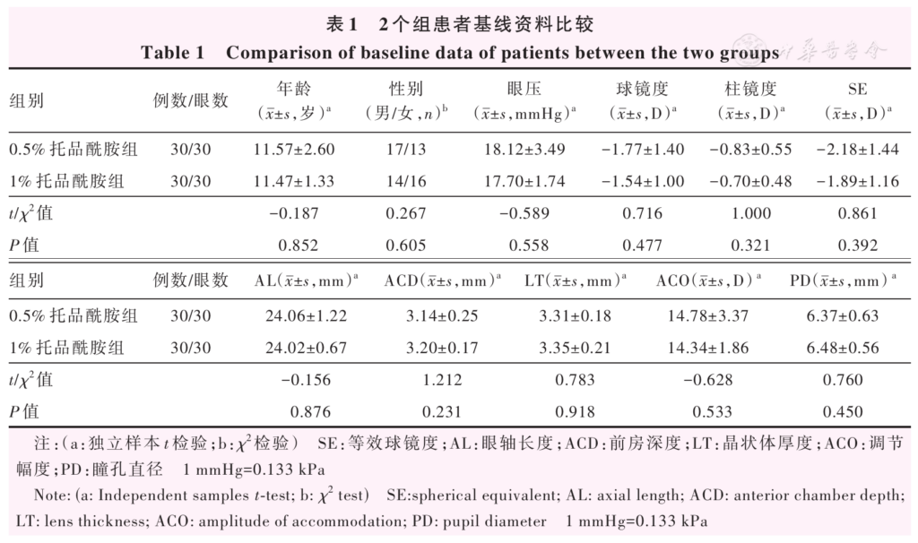
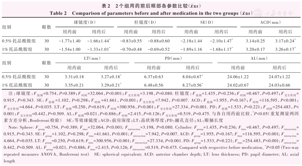
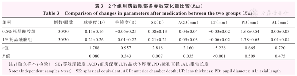
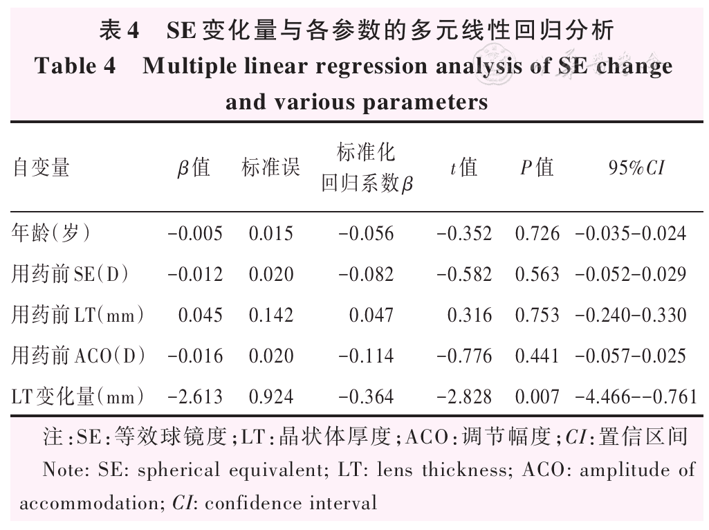
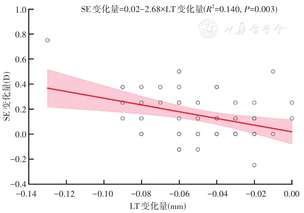
Figure 1 Univariate regression of LT change and SE change LT: lens thickness; SE: spherical equivalent
References click to collapse
[1] Haarman A, Enthoven CA, Tideman J, et al. The complications of myopia: a review and meta‑analysis [J]. Invest Ophthalmol Vis Sci, 2020, 61 (4):49. DOI: 10.1167/iovs.61.4.49.
[2] Fricke TR, Holden BA, Wilson DA, et al. Global cost of correcting vision impairment from uncorrected refractive error [J]. Bull World Health Organ, 2012, 90 (10):728‑738. DOI: 10.2471/BLT.12.104034.
[3] Holden BA, Fricke TR, Wilson DA, et al. Global prevalence of myopia and high myopia and temporal trends from 2000 through 2050 [J]. Ophthalmology, 2016, 123 (5):1036‑1042. DOI: 10.1016/j.ophtha.2016.01.006.
[4] Panke K, Jorova M. Objective refraction status before and after cycloplegia: from childhood to young adulthood [J]. Vision (Basel), 2024, 8 (3):51. DOI: 10.3390/vision8030051.
[5] Gu F, Gao HM, Zheng X, et al. Effect of cycloplegia on refractive error measure in Chinese school students [J]. Ophthalmic Epidemiol, 2022, 29 (6):629‑639. DOI: 10.1080/09286586.2021.1999986.
[6] Yazdani N, Sadeghi R, Momeni‑Moghaddam H, et al. Comparison of cyclopentolate versus tropicamide cycloplegia: a systematic review and meta‑analysis [J]. J Optom, 2018, 11 (3):135‑143. DOI: 10.1016/j.optom.2017.09.001.
[7] Applebaum M, Jaanus SD. Use of diagnostic pharmaceutical agents and incidence of adverse effects [J]. Am J Optom Physiol Opt, 1983, 60 (5):384‑388. DOI: 10.1097/00006324‑198305000‑00007.
[8] Kyei S, Nketsiah AA, Asiedu K, et al. Onset and duration of cycloplegic action of 1% cyclopentolate‑1% tropicamide combination [J]. Afr Health Sci, 2017, 17 (3):923‑932. DOI: 10.4314/ahs.v17i3.36.
[9] North RV, Kelly ME. A review of the uses and adverse effects of topical administration of atropine [J]. Ophthalmic Physiol Opt, 1987, 7 (2):109‑114. DOI: 10.1111/j.1475‑1313.1987.tb01004.x.
[10] Bartlett JD. Administration of and adverse reactions to cycloplegic agents [J]. Am J Optom Physiol Opt, 1978, 55 (4):227‑233. DOI: 10.1097/00006324‑197804000‑00003.
[11] Contreras‑Salinas H, Orozco‑Ceja V, Romero‑López MS, et al. Ocular cyclopentolate: a mini review concerning its benefits and risks [J]. Clin Ophthalmol, 2022, 16:3753‑3762. DOI: 10.2147/OPTH.S388982.
[12] 中华医学会眼科学分会斜视与小儿眼科学组。中国儿童睫状肌麻痹验光及安全用药专家共识 (2019 年)[J]. 中华眼科杂志,2019, 55 (1):7‑12. DOI: 10.3760/cma.j.issn.0412‑4081.2019.01.003.
[13] Pei R, Liu Z, Rong H, et al. A randomized clinical trial using cyclopentolate and tropicamide to compare cycloplegic refraction in Chinese young adults with dark irises [J]. BMC Ophthalmol, 2021, 21 (1):256. DOI: 10.1186/s12886‑021‑02001‑6.
[14] Lin F, Wang Y, Liu Y, et al. The influence of 0.5% tropicamide on anterior segment parameters with CASIA2 in emmetropic, myopic, and hyperopic eyes [J]. Front Physiol, 2022, 13:957097. DOI: 10.3389/fphys.2022.957097.
[15] Liu Y, Shang J, Wang Y, et al. Refractive and ocular biometric characteristics of non‑myopic and pseudomyopic eyes in mild hyperopic Chinese children aged 3‑12 years [J/OL]. Heliyon, 2024, 10 (13):e33987 [2025‑10‑10]. https://pubmed.ncbi.nlm.nih.gov/39055816/. DOI: 10.1016/j.heliyon.2024.e33987.
[16] Manny RE, Hussein M, Scheiman M, et al. Tropicamide (1%): an effective cycloplegic agent for myopic children[J]. Invest Ophthalmol Vis Sci, 2001, 42(8):1728‑1735.
[17] van Minderhout HM, Joosse MV, Grootendorst DC, et al. Eye colour and skin pigmentation as significant factors for refractive outcome and residual accommodation in hypermetropic children: a randomized clinical trial using cyclopentolate 1% and tropicamide 1%[J]. Acta Ophthalmol, 2022, 100(4):454‑461. DOI: 10.1111/aos.15016.
[18] McMullen RJ, Davidson MG, Gilger BC. The effect of 1% tropicamide induced mydriasis and cycloplegia on spherical refraction of the adult horse[J]. Vet Ophthalmol, 2014, 17(2):120‑125. DOI: 10.1111/vop.12055.
[19] Yuan Y, Zhang Z, Zhu J, et al. Responses of the ocular anterior segment and refraction to 0.5% tropicamide in Chinese school‑aged children of myopia, emmetropia, and hyperopia[J]. J Ophthalmol, 2015, 2015:612728. DOI: 10.1155/2015/612728.
[20] Vinay PG, Bhat S, Mathews MM. A comparison of the mydriatic efficacy of tropicamide and phenylephrine eye drops administered as a vaporized spray in one eye and conventional drop instillation in the other eye in an Indian pediatric cohort[J]. J Curr Ophthalmol, 2022, 34(4):448‑451. DOI: 10.4103/joco.joco_127_22.
[21] Wong CY, Fan DS, Yu CB, et al. Topical mydriatic and cycloplegic spray for Chinese children[J]. J Pediatr Ophthalmol Strabismus, 2003, 40(6):349‑352. DOI: 10.3928/0191‑3913‑20031101‑08.
[22] Alghamdi WM, Alrasheed SH, Nair V, et al. Effects of cyclopentolate hydrochloride dosage on anterior segment parameters in young adults (measured with Pentacam)[J]. Clin Ophthalmol, 2021, 15:891‑898. DOI: 10.2147/OPTH.S291991.
[23] Pan CW, Zhang O, Hu DN, et al. Iris color and lens thickness in Chinese teenagers[J]. Transl Vis Sci Technol, 2018, 7(5):25. DOI: 10.1167/tvst.7.5.25.
[24] Yassa ET, Ünlü C. Comparison of autorefraction and photorefraction with and without cycloplegia using 1% tropicamide in preschool children[J]. J Ophthalmol, 2019, 2019:1487013. DOI: 10.1155/2019/1487013.
[25] Zhang J, Ni Y, Li P, et al. Anterior segment biometry with phenylephrine and tropicamide during accommodation imaged with ultralong scan depth optical coherence tomography[J]. J Ophthalmol, 2019, 2019:6827215. DOI: 10.1155/2019/6827215.
[26] Lockett‑Ruiz V, Navarro R, López‑Gil N. Intracapsular accommodation mechanism in terms of lens curvature gradient[J]. Ophthalmic Physiol Opt, 2024, 44(2):334‑346. DOI: 10.1111/opo.13271.
[27] Pumphrey SA, Moon Y, Francis JM, et al. Duration of mydriasis produced by 0.5% and 1% tropicamide in Sprague‑Dawley rats[J]. J Am Assoc Lab Anim Sci, 2021, 60(5):576‑581. DOI: 10.30802/AALAS‑JAALAS‑21‑000013.
[28] Momeni‑Moghaddam H, Maddah N, Wolffsohn JS, et al. The effect of cycloplegia on the ocular biometric and anterior segment parameters: a cross‑sectional study[J]. Ophthalmol Ther, 2019, 8(3):387‑395. DOI: 10.1007/s40123‑019‑0187‑5.