Authors:Wu Feng, Li Man, Liu Xiaona
Corresponding author:Liu Xiaona, Email:liuxiaona128@163.com
Published:2026-08-10
DOI:10.3760/cma.j.cn115989-20240501-00121
ABSTRACT
This study reported the diagnosis and treatment process of a 74-year-old male patient with left eye fungal corneal ulcer caused by Sidea alba induced by ocular trauma. The patient presented with red eye, ocular pain and blurred vision after left eye trauma, and the symptoms were not improved after more than one month of topical eye drop treatment in a local hospital. Ocular examination, laser scanning confocal microscopy, fungal fluorescence staining and mass spectrometry identification were performed after admission, and the pathogen was confirmed as Sidea alba. Initial conservative treatment with topical and systemic antifungal drugs failed to control the disease progression. Left corneal lesion resection was performed timely, and the regimens of antifungal, anti-inflammatory and corneal repair drugs were dynamically adjusted according to postoperative ocular recovery. The patient’s ocular symptoms were gradually relieved, the corneal ulcer decreased in size and healed gradually after operation, and complete cure was achieved during long-term follow-up.
KEYWORDS:
Corneal ulcer; Fungal keratitis; Sidea alba; Ocular trauma; Antifungal agents
COPYRIGHTS:
Copyright by Chinese Medical Association
No content published by the journals of Chinese Medical Association may be reproduced or abridged without authorization. Please do not use or copy the layout and design of the journals without permission.
All articles published represent the opinions of the authors, and do not reflect the official policy of the Chinese Medical Association or the Editorial Board, unless this is clearly specified.
Authors Info & Affiliations
Wu Feng
Jinan Mingshui Eye Hospital Laboratory, Jinan 250200, China
Li Man
Jinan Mingshui Eye Hospital Ophthalmology, Jinan 250200, China
Liu Xiaona
Jinan Mingshui Eye Hospital Laboratory, Jinan 250200, China
Figures & Tables
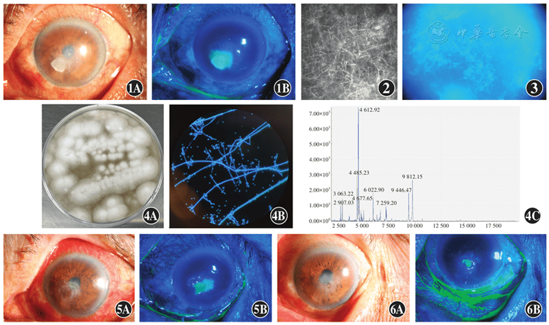
Figure 1 Preoperative slit-lamp microscope images A gray-white opaque infiltrative lesion of approximately 3 mm×4 mm was observed at the inferonasal central cornea of the left eye, with crust-like secretions on the surface, peripheral corneal pseudopodia, stromal edema, Descemet membrane folds, and punctate and patchy gray-white keratic precipitates A: Diffuse illumination B: Cobalt blue light.
Figure 2 Preoperative laser scanning confocal microscopy images A large number of dense hypha-like high-reflective signals were detected in the corneal ulcer area of the left eye. The endothelial structure around the lesion was unclear, with numerous punctate intense reflective spots
Figure 3 Fungal fluorescence staining of corneal scraping (calcofluor white staining, ×400) Hypha-like structures were visible
Figure 4 Culture morphology, staining results and mass spectrometry identification of corneal scraping isolates A: White flocculent colonies of the culture B: Structure of hyphae and spores seen under the microscope after fluorescent staining of the culture (× 1 000) C: Mass spectrometer identification results
Figure 5 Slit-lamp microscope images at 3 days after operation The opacity of the inferonasal central corneal ulcer in the left eye was alleviated, with a small amount of mucinous and white secretions on the surface, and partial repair of the corneal epithelium around the ulcer A: Diffuse illumination B: Cobalt blue light
Figure 6 Slit-lamp microscope images at 3 weeks after operation The corneal ulcer showed clear boundary with reduced size and no abnormal secretions on the surface A: Diffuse illumination B: Cobalt blue light
References click to collapse
[1] Priyadarshini SR, Soni T, Sahu SK, et al. Acremonium keratitis: risk factors, clinical characteristics, management, and outcome in 65 cases[J]. Indian J Ophthalmol,2022, 70(10):3522-3527. DOI: 10.4103/ijo.IJO_659_22.
[2] GargP, RoyA, RoyS. Update on fungal keratitis[J]. Curr Opin Ophthalmol, 2016,27(4):333-339.DOI:10.1097/ICU.0000000000000272.
[3] Tsang C, Chan JFW, Pong W, et al. Cutaneous hyalohyphomycosis due to Parengyodontium album gen. et comb. nov[J]. Med Mycol, 2016, 54(7):699-713. DOI: 10.1093/mmy/myw025.
[4] Chellappan S, Jasmin C, Basheer SM, et al. Production, purification and partial characterization of a novel protease from marine Engyodontium album BTMFS10 under solid state fermentation[J]. Process Biochemistry, 2006, 41(4):956-961.
[5] Šimonovičová A, Gódyová M, Kunert J. Engyodontium album, a new species of microscopic fungi for Slovakia and its keratinolytic activity[J]. Biologia (Bratisl), 2004, 59(1):17-18.
[6] Macêdo DPC, Neves RP. de Souza‐Motta CM, et al. Engyodontium album fungaemia: the first reported case[J]. Braz J Microbiol, 2007, 38:110-112.
[7] Yao Q, Wang J, Zhang X, et al. Cytotoxic polyketides from the deep‐sea‐derived fungus Engyodontium album DFFSCS021[J]. Mar Drugs, 2014, 12(12):5902-5915. DOI: 10.3390/md12125902.
[8] Kim HS, Park IY, Park YJ, et al. A novel dihydroxanthenone, AGI‐B4 with inhibition of VEGF‐induced endothelial cell growth[J]. J Antibiot (Tokyo), 2002, 55(7):669-672. DOI: 10.7164/antibiotics.55.669.
[9] KanegaeH, TominoN, NakamuraY, et al. Parengyodontium album isolated from cutaneous lesions of a Pacific white‐sided dolphin (Lagenorhynchus obliquidens) during treatment for paracoccidioidomycosis ceti[J]. Mycopathologia, 2020, 185(6):1021-1031. DOI: 10.1007/s11046‐020 00484‐3.
[10]Mohamed N, Ding CH, Wahab AA, et al. Isolation of an obscure fungus, Parengyodontium album, from the blood of a severely neutropenic patient: the first reported case from Malaysia[J]. J Infect Dev Ctries, 2022, 16(10):1668-1670. DOI: 10.3855/jidc.17016.
[11]Thamke DC, Mendiratta DK, Dhabarde A, et al. Mycotic keratitis due to Engyodontium album: first case report from India[J]. Indian J Med Microbiol, 2015, 33(2):303-304. DOI: 10.4103/0255‐0857.154890.