Authors:Yuan Chaofeng, Meng Jia, Shao Jingzhi, Ma Chengxia, Huang Xuetao,Du Liping
Corresponding author: Du Liping, Email:1307438431@qq.com
Published:2026-08-10
DOI:10.3760/cma.j.cn115989-20260120-00036
ABSTRACT
Objective To explore the effects of intraoperative anterior chamber dexamethasone injection on inflammatory response and visual function in patients with uveitis complicated with cataract and closed pupil membrane.
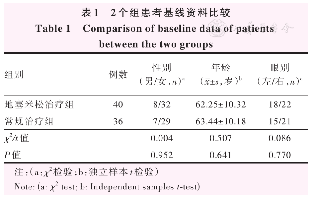
Methods A cohort study was conducted. A total of 76 patients (76 eyes) with cataracts complicated by uveitis who were treated at the First Affiliated Hospital of Zhengzhou University from June 2024 to May 2025 were enrolled. All patients underwent routine treatment combined with phacoemulsification and intraocular lens implantation. Intraoperatively, dexamethasone was injected into the anterior chamber in 40 cases (40 eyes), which were assigned to the dexamethasone treatment group, while the remaining 36 cases (36 eyes) were assigned to the routine treatment group. Visual acuity was measured at 1 week and 1 month postoperatively, and the visual function improvement rates were calculated preoperatively. The incidences of complications, including corneal edema, anterior chamber exudation, and anterior chamber flare, were recorded. Serum levels of interleukin-6 (IL-6), C-reactive protein (CRP), and tumor necrosis factor-α (TNF-α) were determined by enzyme-linked immunosorbent assay to evaluate the systemic inflammatory response. This study complied with the Declaration of Helsinki. The study protocol was approved by the Ethics Committee of the First Affiliated Hospital of Zhengzhou University (No. 2025-KY-1151). Both the patients themselves or their guardians gave informed consent to the study.
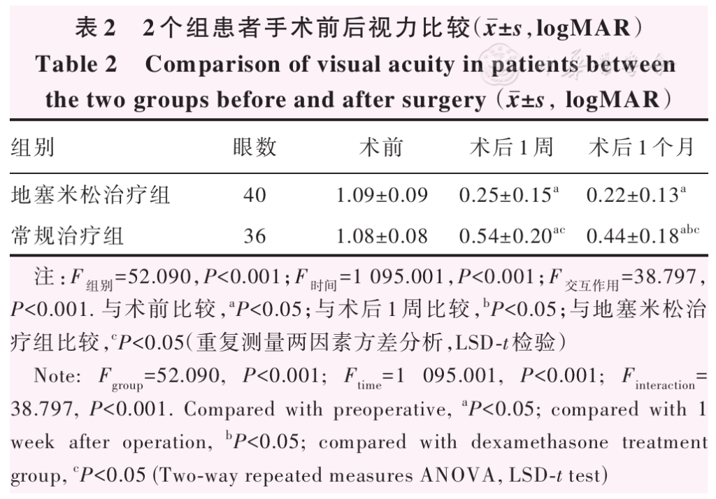
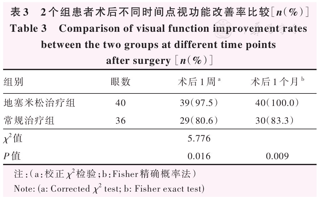
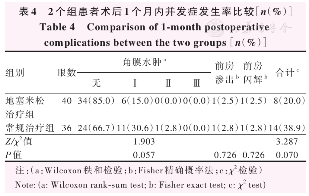
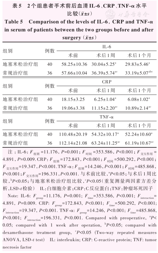
Results The preoperative, 1-week postoperative and 1-month postoperative visual acuity were 1.09±0.09, 0.25±0.15 and 0.22±0.13 in the dexamethasone treatment group, respectively, and 1.08±0.08, 0.54±0.20, and 0.44±0.18 in the routine treatment group, respectively, with statistically significant overall differences ( F group=52.090, P<0.001; F time=1 095.001, P<0.001). The 1-week postoperative and 1-month postoperative visual acuity were significantly better in the dexamethasone treatment group than in the routine treatment group (both P<0.05). In the dexamethasone treatment group and routine treatment group, the visual acuity at 1 week and 1 month postoperatively were significantly better than the preoperative level, and the visual acuity at 1 month postoperatively was significantly better than that at 1 week postoperatively in the routine treatment group (all P<0.05). The 1-week postoperative and 1-month postoperative visual function improvement rates were significantly higher in the dexamethasone treatment group than in the routine treatment group (both P<0.05) and no statistically significant differences were observed in the incidences of corneal edema, anterior chamber exudation, anterior chamber flare, and the total complication rate within 1 month postoperatively between the two groups (all P>0.05). The 1-week postoperative and 1-month postoperative serum levels of IL-6, CRP, and TNF-α were significantly lower in the dexamethasone treatment group than in the routine treatment group (all P<0.05). In the dexamethasone treatment group and routine treatment group, the serum levels of IL-6, CRP, and TNF-α at 1 week and 1 month postoperatively were significantly lower than the preoperative levels, and the serum IL-6 level at 1 month postoperatively was significantly lower than that at 1 week postoperatively in the routine treatment group ( P<0.05).
Conclusions Intraoperative anterior chamber injection of dexamethasone alleviates postoperative inflammatory responses, improves visual function for patients with uveitis complicated by cataracts and pupillary membrane occlusion, and is safe and reliable.
KEYWORDS:
Dexamethasone;Cataract;Uveitis;Pupillary membrane closure;Inflammatory reactions;Visual function
COPYRIGHTS:
Copyright by Chinese Medical Association
No content published by the journals of Chinese Medical Association may be reproduced or abridged without authorization. Please do not use or copy the layout and design of the journals without permission.
All articles published represent the opinions of the authors, and do not reflect the official policy of the Chinese Medical Association or the Editorial Board, unless this is clearly specified.
Authors Info & Affiliations
Yuan Chaofeng
Department of Ophtalmology, The First Affiliated Hospital of Zhengzhou University, Henan Provincial Eye Hospital, Zhengzhou 450000, China
Meng Jia
Department of Ophtalmology, The First Affiliated Hospital of Zhengzhou University, Henan Provincial Eye Hospital, Zhengzhou 450000, China
Shao Jingzhi
Department of Ophtalmology, The First Affiliated Hospital of Zhengzhou University, Henan Provincial Eye Hospital, Zhengzhou 450000, China
Ma Chengxia
Department of Ophtalmology, The First Affiliated Hospital of Zhengzhou University, Henan Provincial Eye Hospital, Zhengzhou 450000, China
Huang Xuetao
Department of Ophtalmology, The First Affiliated Hospital of Zhengzhou University, Henan Provincial Eye Hospital, Zhengzhou 450000, China
Du Liping
Department of Ophtalmology, The First Affiliated Hospital of Zhengzhou University, Henan Provincial Eye Hospital, Zhengzhou 450000, China
Figures & Tables
References click to collapse
[1] Agrawal R, Testi I, Bodaghi B, et al. Collaborative ocular tuberculosis study consensus guidelines on the management of tubercular uveitis‑report 2: guidelines for initiating antitubercular therapy in anterior uveitis, intermediate uveitis, panuveitis, and retinal vasculitis [J]. Ophthalmology, 2021, 128 (2):277-287. DOI: 10.1016/j.ophtha.2020.06.052.
[2] Abdalla Elsayed M, Kozak I. Pharmacologically induced uveitis [J]. Surv Ophthalmol, 2021, 66 (5):781-801. DOI: 10.1016/j.survophthal.2021.01.003.
[3] Bagheri M, Ahoor MH, Jafari A, et al. Pattern of uveitis in Iran: a systematic review [J]. J Ophthalmic Vis Res, 2021, 16 (1):93-102. DOI: 10.18502/jovr.v16i1.8255.
[4] Oh BL, Lee JS, Lee EY, et al. Incidence and risk factors for blindness in uveitis: a nationwide cohort study from 2002 to 2013 [J]. Ocul Immunol Inflamm, 2021, 29 (6):1040-1044. DOI: 10.1080/09273948.2020.1746352.
[5] Nguyen QD, Anesi SD, Chexal S, et al. Management of repository corticotropin injection therapy for non‑infectious uveitis: a Delphi study [J]. Acta Ophthalmol, 2021, 99 (6):669-678. DOI: 10.1111/aos.14702.
[6] Gagnon NA, Hartley C, Gilger BC. Efficacy and safety of suprachoroidal triamcinolone injection in horses with poorly responsive equine recurrent uveitis [J]. Vet Ophthalmol, 2021, 24 (3):308-312. DOI: 10.1111/vop.12887.
[7] Halim J, Westcott F, Cascone N, et al. Risk factors associated with post‑operative uveitis after cataract surgery: a retrospective case‑control study [J]. Eye (Lond), 2022, 36 (1):198-205. DOI: 10.1038/s41433‑021‑01486‑1.
[8] Accorinti M, Saturno MC, Paroli MP, et al. Uveitis‑glaucoma‑hyphema syndrome: clinical features and differential diagnosis [J]. Ocul Immunol Inflamm, 2022, 30 (6):1408-1413. DOI: 10.1080/09273948.2021.1881563.
[9] Smith JR, Matthews JM, Conrad D, et al. Recommendations for the management of childhood juvenile idiopathic arthritis‑type chronic anterior uveitis [J]. Clin Exp Ophthalmol, 2021, 49 (1):38-45. DOI: 10.1111/ceo.13856.
[10] De Maria M, Coassin M, Iannetta D, et al. Laser flare and cell photometry to measure inflammation after cataract surgery: a tool to predict the risk of cystoid macular edema [J]. Int Ophthalmol, 2021, 41 (6):2293-2300. DOI: 10.1007/s10792‑021‑01779‑0.
[11] Dubashynskaya NV, Bokatyi AN, Skorik YA. Dexamethasone conjugates: synthetic approaches and medical prospects [J]. Biomedicines, 2021, 9 (4):341. DOI: 10.3390/biomedicines9040341.
[12] Wilkins CS, Sobol EK, Lema GM, et al. Intravitreal dexamethasone insert in diabetic macular edema super‑refractory to anti‑vascular endothelial growth factor therapy [J]. Eur J Ophthalmol, 2022, 32 (5):NP37‑NP41. DOI: 10.1177/11206721211004391.
[13] Bodaghi B, Brézin AP, Weber M, et al. Real‑life efficacy, safety, and use of dexamethasone intravitreal implant in posterior segment inflammation due to non‑infectious uveitis (LOUVRE 2 Study)[J]. Ophthalmol Ther, 2022, 11 (5):1775‑1792. DOI: 10.1007/s40123‑022‑00525‑8.
[14] Gupta G, Ram J, Gupta V, et al. Efficacy of intravitreal dexamethasone implant in patients of uveitis undergoing cataract surgery [J]. Ocul Immunol Inflamm, 2019, 27 (8):1330‑1338. DOI: 10.1080/09273948.2018.1524498.
[15] 王宁利,李仕明,李文生。循证眼科学 [M]. 北京:人民卫生出版社,2020:141‑146.
[16] 黎晓新。实用眼科学 (第 4 版)[M]. 北京:人民卫生出版社,2023:112‑116.
[17] Hsu AY, Kuo HT, Lin CJ, et al. Cataract development among pediatric patients with uveitis [J/OL]. JAMA Netw Open, 2024, 7 (7):e2419366 [2026‑01‑02]. https://pubmed.ncbi.nlm.nih.gov/38949811/. DOI: 10.1001/jamanetworkopen.2024.19366.
[18] 李学佳,张开朗,陈思思,等. lncRNA 5033413D16Rik 对实验性自身免疫性葡萄膜炎 Th17 细胞活性的抑制作用 [J]. 中华实验眼科杂志,2024, 42 (11):983‑990. DOI: 10.3760/cma.j.cn115989‑20230220‑00056.
Li XJ, Zhang KL, Chen SS, et al. Inhibitory effect of lncRNA 5033413D16Rik on the activity of T helper 17 cells in experimental autoimmune uveitis [J]. Chin J Exp Ophthalmol, 2024, 42 (11):983‑990. DOI: 10.3760/cma.j.cn115989‑20230220‑00056.
[19] Shekhar M, Chaudhary S, Pai A, et al. Visual outcomes and risk factors of uveitis cataract eyes after phacoemulsification and manual small‑incision cataract surgery [J]. J Cataract Refract Surg, 2024, 50 (3):257‑263. DOI: 10.1097/j.jcrs.0000000000001354.
[20] Yu X, Duan R, Jiang L, et al. Interleukin‑6 in non‑infectious uveitis: biology, experimental evidence and treatment strategies [J]. Biochem Pharmacol, 2024, 230 (Pt 2):116605. DOI: 10.1016/j.bcp.2024.116605.
[21] Erdoğan S, Sorgun MH, Kuzu Mumcu M, et al. Nutritional status in patients with Neuro‑Behçet’s disease [J]. Turk J Med Sci, 2021, 51 (4):1682‑1688. DOI: 10.3906/sag‑2009‑235.
[22] Bernal‑Morales C, Ramanan AV, Pavesio C. Use of immunomodulators in non‑infectious uveitis: lights and shadows [J]. Eye (Lond), 2024, 38 (17):3231‑3242. DOI: 10.1038/s41433‑024‑03294‑9.
[23] Chychko L, Son HS, Friedrich M, et al. Molecular changes in aqueous humor associated with inflammation following cataract surgery in patients with Fuchs’ endothelial corneal dystrophy [J]. Ophthalmol Ther, 2025, 14 (1):197‑209. DOI: 10.1007/s40123‑024‑01072‑0.
[24] Agarwal S, Gupta PC, Shamim MA, et al. Outcomes of pediatric cataract surgery: intraoperative triamcinolone injection versus dexamethasone injection [J]. Indian J Ophthalmol, 2026, 74 (1):53‑58. DOI: 10.4103/IJO.IJO_2600_24.
[25] Li YT, Cui XX, Yang XT, et al. Utilizing dexamethasone intravitreal implant to control postoperative inflammation in refractory uveitis undergoing cataract surgery [J]. Int J Ophthalmol, 2021, 14 (2):317‑322. DOI: 10.18240/ijo.2021.02.21.
[26] Nguyen BM, Do CL, Boydstun IP, et al. Iris atrophy following intracameral dexamethasone injection: a report of two pediatric cases [J]. J AAPOS, 2024, 28 (3):103901. DOI: 10.1016/j.jaapos.2024.103901.