Authors:Miao Zhuang, Mo Jing, Lu Qi, Li Qinglan
Corresponding author: Mo Jing, Email:mojing@aierchina.com
Published:2026-08-10
DOI:10.3760/cma.j.cn115989-20250423-00132
ABSTRACT
Objective To analyze the dynamic changes of multiple corneal parameters and the safety of femtosecond laser small-incision lenticule extraction combined with corneal cross-linking (SMILE Xtra) 12 months postoperatively in patients with moderate-to-high risk of corneal ectasia.
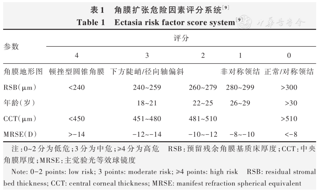
Methods A prospective self-control study was conducted. A total of 31 myopic patients (54 eyes) who underwent SMILE Xtra at Aier Eye Hospital (East of Chengdu) from May 2023 to February 2024 were enrolled. All patients were classified as moderate to high risk according to the Randleman Ectasia Risk Score System (ERSS). Uncorrected distance visual acuity (UDVA), manifest refraction spherical equivalent (MRSE), intraocular pressure (IOP), maximum keratometry (Kmax), mean keratometry (Kmean), corneal morphological indices including central keratoconus index (CKI), index of highest decentration (IHD), index of surface variance (ISV), and index of vertical asymmetry (IVA), corneal biomechanical parameters including stiffness parameter at first applanation (SP-A1), stress strain index (SSI), and integrated radius (IR), corneal densitometry (CD) were recorded preoperatively and at 1, 3, 6, and 12 months postoperatively, and complications were also recorded. This study followed the Declaration of Helsinki. The study protocol was approved by the Ethics Committee of Aier Eye Hospital (East of Chengdu) (No. DQAIER202303001). All patients signed the informed consent form.
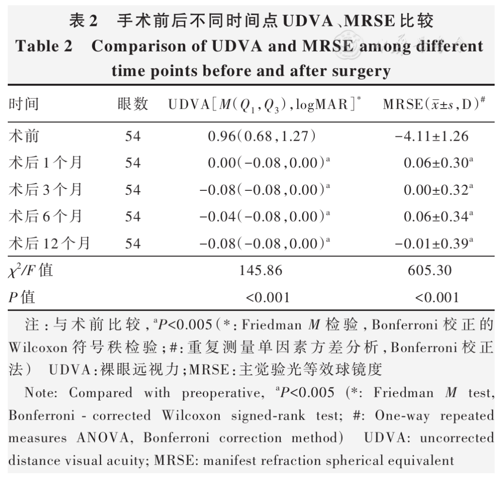
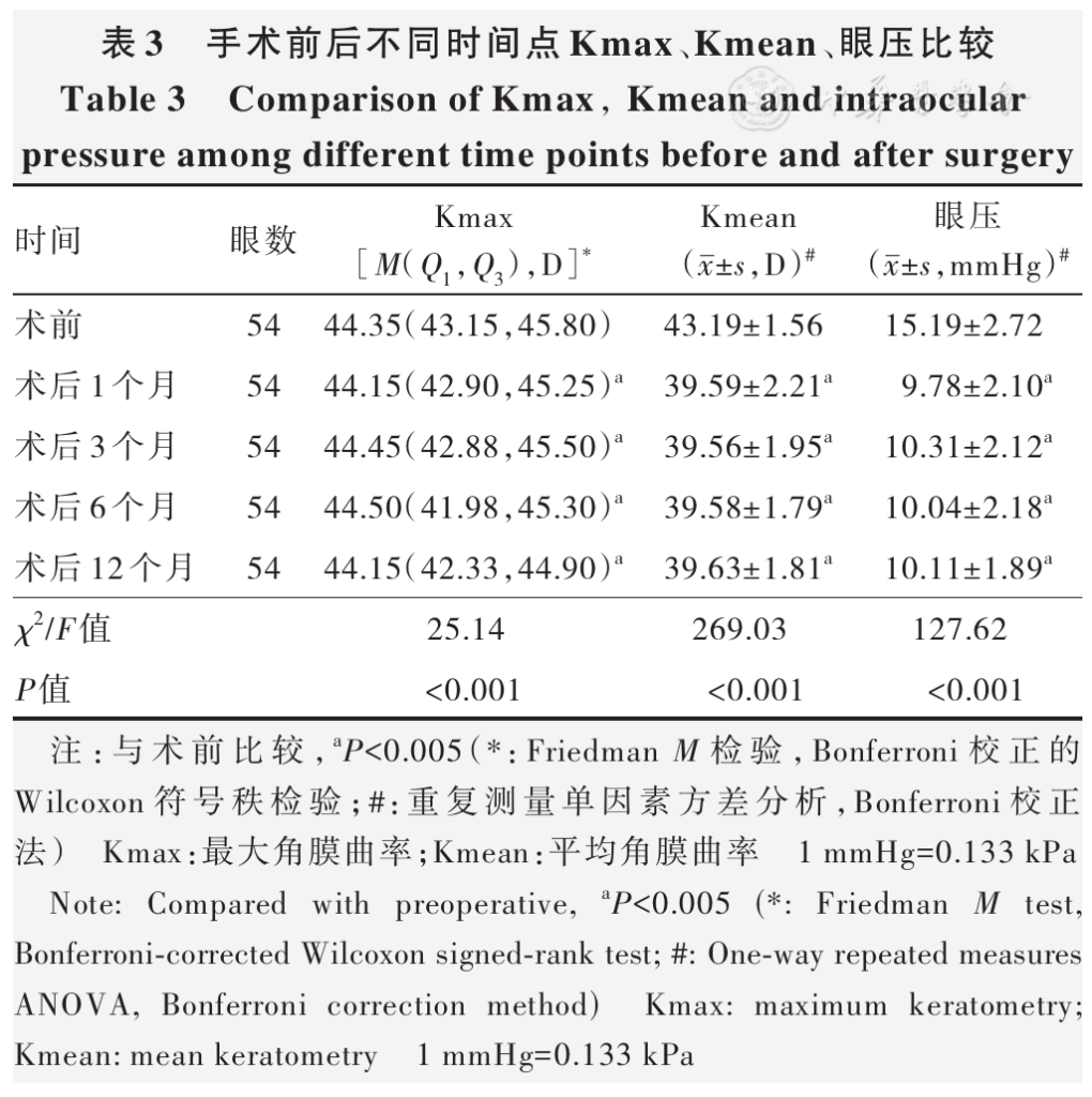
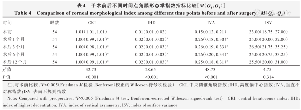
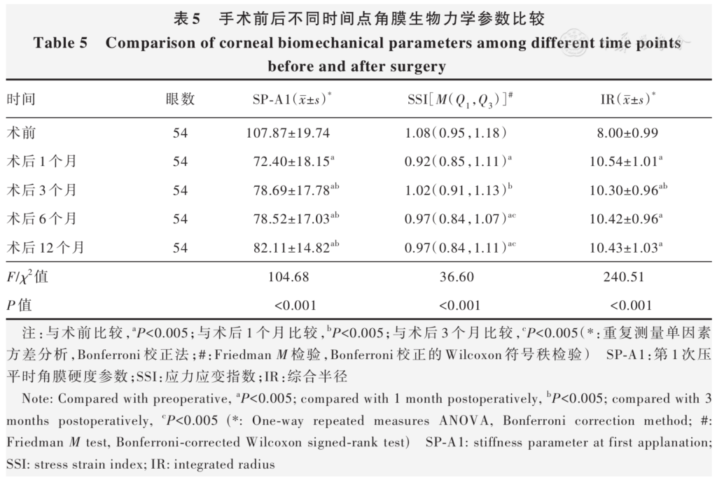
Results UDVA, absolute value of MRSE, Kmax, Kmean, IOP, CKI, and SP-A1 at different postoperative time points were significantly decreased compared with preoperative, while IHD, IVA, and IR were significantly increased (all P<0.005). SP-A1 at 3, 6, and 12 months postoperatively was higher than that at 1 month postoperatively, SSI at 1, 6, and 12 months postoperatively was significantly decreased compared with preoperative, SSI at 3 months postoperatively was higher than that at 1, 6, and 12 months postoperatively, and IR at 3 months was significantly decreased compared with that at 1 month postoperatively (all P<0.005). There were no statistically significant differences in pairwise comparisons of UDVA, MRSE, Kmax, Kmean, IOP, CKI, IHD, and IVA at any postoperative time points (all P>0.005). No statistically significant difference was found in ISV across different time points ( χ 2=4.75, P=0.314). Preoperative and postoperative CD at 1, 3, 6, and 12 months were (16.92±1.28), (17.89±1.76), (17.83±1.80), (17.45±1.90), and (17.30±1.70) grayscale units ( GSU), respectively, with a statistically significant overall difference ( F=9.57, P<0.001). Specifically, CD at 1 and 3 months postoperatively was significantly increased compared with preoperative (both P<0.001); CD at 6 and 12 months after surgery recovered to preoperative baseline level, with no statistically significant differences (both P>0.005). At 1 month postoperatively, grade 0.5 haze was observed in 4 eyes (7.4%) and grade 1 haze in 9 eyes (16.7%); at 3 months postoperatively, grade 0.5 haze was observed in 8 eyes (14.8%) and grade 1 haze in 3 eyes (5.6%). All haze resolved within 6 months after treatment.
Conclusions SMILE Xtra demonstrates favorable efficacy, stability, and safety in correcting ametropia among patients with moderate to high risk ERSS within 12 months postoperatively. Corneal biomechanical properties at 3 months postoperatively were enhanced compared with those at 1 month postoperatively. Nevertheless, SSI variability and haze occurrence postoperatively demand clinical attention.
KEYWORDS:
Myopia;Small incision lenticule extraction;Corneal cross-linking;Corneal biomechanics;Corneal morphology;Corneal densitometry
COPYRIGHTS:
Copyright by Chinese Medical Association
No content published by the journals of Chinese Medical Association may be reproduced or abridged without authorization. Please do not use or copy the layout and design of the journals without permission.
All articles published represent the opinions of the authors, and do not reflect the official policy of the Chinese Medical Association or the Editorial Board, unless this is clearly specified.
Authors Info & Affiliations
Miao Zhuang
Aier Eye Hospital (East of Chengdu), Chengdu 610056, China
Mo Jing
Aier Eye Hospital (East of Chengdu), Chengdu 610056, China
Lu Qi
Aier Eye Hospital (East of Chengdu), Chengdu 610056, China
Li Qinglan
Aier Eye Hospital (East of Chengdu), Chengdu 610056, China
Figures & Tables
References click to collapse
[1] Liang J, Pu Y, Chen J, et al. Global prevalence, trend and projection of myopia in children and adolescents from 1990 to 2050: a comprehensive systematic review and meta‑analysis [J]. Br J Ophthalmol, 2025, 109 (3):362-371. DOI: 10.1136/bjo‑2024‑325427.
[2] 中华医学会眼科学分会眼视光学组。我国飞秒激光小切口角膜基质透镜取出手术规范专家共识 (2018 年)[J]. 中华眼科杂志,2018,54 (10):729-736. DOI: 10.3760/cma.j.issn.0412‑4081.2018.10.003.
Ophthalmic Optometry Group of Ophthalmology Branch of the Chinese Medical Association. Expert consensus on standards for small incision lenticule extraction (SMILE) in China (2018)[J]. Chin J Ophthalmol, 2018, 54 (10):729-736. DOI: 10.3760/cma.j.issn.0412‑4081.2018.10.003.
[3] Wang Y, Cui C, Li Z, et al. Corneal ectasia 6.5 months after small‑incision lenticule extraction [J]. J Cataract Refract Surg, 2015, 41 (5):1100-1106. DOI: 10.1016/j.jcrs.2015.04.001.
[4] Moshirfar M, Tukan AN, Bundogji N, et al. Ectasia after corneal refractive surgery: a systematic review [J]. Ophthalmol Ther, 2021, 10 (4):753-776. DOI: 10.1007/s40123‑021‑00383‑w.
[5] 中华医学会眼科学分会眼视光学组,中国医师协会眼科医师分会眼视光专业委员会,中国医师协会眼科医师分会屈光手术专业委员会。中国角膜交联术治疗扩张性角膜疾病专家共识 (2023)[J]. 中华眼视光学与视觉科学杂志,2023, 25 (12):881-888. DOI: 10.3760/cma.j.cn115909‑20231106‑00176.
Optometry Group of Ophthalmology Branch of Chinese Medical Association, Ophthalmology and Optometry Committee of Ophthalmologist Association of Chinese Doctor Association, Refractive Surgery Group of Ophthalmologist Association of Chinese Doctor Association. Consensus on the corneal cross‑linking in the treatment of corneal ectasia disease in China (2023)[J]. Chin J Optom Ophthalmol Vis Sci, 2023, 25 (12):881-888. DOI: 10.3760/cma.j.cn115909‑20231106‑00176.
[6] Papachristoforou N, Ueno A, Ledwos K, et al. A review of keratoconus cross‑linking treatment methods [J]. J Clin Med, 2025, 14 (5):1702. DOI: 10.3390/jcm14051702.
[7] Brar S, Sriganesh S, Sute SS, et al. Comparison of long‑term outcomes and refractive stability following SMILE versus SMILE combined with accelerated cross‑linking (SMILE XTRA)[J]. J Ophthalmol, 2022, 2022:4319785. DOI: 10.1155/2022/4319785.
[8] 莫菲,李莹。屈光手术联合预防性角膜胶原交联矫正屈光不正的疗效及安全性研究进展 [J]. 中华实验眼科杂志,2024,42 (4):376-385. DOI: 10.3760/cma.j.cn115989‑20220624‑00295.
Mo F, Li Y. Efficacy and safety of refractive surgery combined with prophylactic collagen cross‑linking for ametropia [J]. Chin J Exp Ophthalmol, 2024, 42 (4):376-385. DOI: 10.3760/cma.j.cn115989‑20220624‑00295.
[9] Randleman JB, Woodward M, Lynn MJ, et al. Risk assessment for ectasia after corneal refractive surgery [J]. Ophthalmology, 2008, 115 (1):37-50. DOI: 10.1016/j.ophtha.2007.03.073.
[10] Maharana PK, Dubey A, Jhanji V, et al. Management of advanced corneal ectasias [J]. Br J Ophthalmol, 2016, 100 (1):34-40. DOI: 10.1136/bjophthalmol‑2015‑307059.
[11] Raiskup F, Spoerl E. Corneal crosslinking with riboflavin and ultraviolet A. Part II. Clinical indications and results [J]. Ocul Surf, 2013, 11 (2):93-108. DOI: 10.1016/j.jtos.2013.01.003.
[12] 张睿倩,付梦军,王锐,等.SMILE 联合角膜胶原交联术后角膜形态学变化 [J]. 国际眼科杂志,2020, 20 (2):290-293. DOI: 10.3980/j.issn.1672‑5123.2020.2.22.
Zhang RQ, Fu MJ, Wang R, et al. Clinical study on the morphological changes of cornea after SMILE combined with corneal collagen cross‑linking [J]. Int Eye Sci, 2020, 20 (2):290-293. DOI: 10.3980/j.issn.1672‑5123.2020.2.22.
[13] Ganesh S, Brar S. Clinical outcomes of small incision lenticule extraction with accelerated cross‑linking (ReLEx SMILE Xtra) in patients with thin corneas and borderline topography [J]. J Ophthalmol, 2015, 2015:263412. DOI: 10.1155/2015/263412.
[14] Ng AL, Chan TC, Cheng GP, et al. Comparison of the early clinical outcomes between combined small‑incision lenticule extraction and collagen cross‑linking versus SMILE for myopia [J]. J Ophthalmol, 2016, 2016:2672980. DOI: 10.1155/2016/2672980.
[15] Sánchez‑González JM, Rocha‑de‑Lossada C, Borroni D, et al. Prophylactic corneal crosslinking in myopic small‑incision lenticule extraction‑ long‑term visual and refractive outcomes [J]. Indian J Ophthalmol, 2022, 70 (1):73-78. DOI: 10.4103/ijo.IJO_810_21.
[16] 蒋莎,雷晓华,谭维娜,等.SMILE 联合 CXL 与 SMILE 矫正近视眼术后早期疗效和角膜生物力学变化比较 [J]. 中华实验眼科杂志,2021,39 (5):430-438. DOI: 10.3760/cma.j.cn115989‑20191208‑00529.
Jiang S, Lei XH, Tan WN, et al. Evaluation and comparison of early outcome and corneal biomechanical changes between SMILE‑corneal collagen cross linking and SMILE for myopic eyes [J]. Chin J Exp Ophthalmol, 2021, 39 (5):430-438. DOI: 10.3760/cma.j.cn115989‑20191208‑00529.
[17] Piyacomn Y, Kasetsuwan N, Puangsricharern V, et al. Topometric indices and corneal densitometry change after corneal refractive surgery combined with simultaneous collagen crosslinking [J]. Clin Ophthalmol, 2019, 13:1927-1933. DOI: 10.2147/OPTH.S225909.
[18] Shen Y, Zhao J, Yao P, et al. Changes in corneal deformation parameters after lenticule creation and extraction during small incision lenticule extraction (SMILE) procedure [J/OL]. PLoS One, 2014, 9 (8):e103893 [2025-07-12]. https://pubmed.ncbi.nlm.nih.gov/25121508/. DOI: 10.1371/journal.pone.0103893.
[19] Cao K, Liu L, Yu T, et al. Changes in corneal biomechanics during small‑incision lenticule extraction (SMILE) and femtosecond‑assisted laser in situ keratomileusis (FS‑LASIK)[J]. Lasers Med Sci, 2020, 35 (3):599-609. DOI: 10.1007/s10103‑019‑02854‑w.
[20] Mencucci R, Marini M, Paladini I, et al. Effects of riboflavin/UVA corneal cross‑linking on keratocytes and collagen fibres in human cornea [J]. Clin Exp Ophthalmol, 2010, 38 (1):49-56. DOI: 10.1111/j.1442‑9071.2010.02207.x.
[21] Eliasy A, Chen KJ, Vinciguerra R, et al. Determination of corneal biomechanical behavior in‑vivo for healthy eyes using CorVis ST tonometry: stress‑strain index [J]. Front Bioeng Biotechnol, 2019, 7:105. DOI: 10.3389/fbioe.2019.00105.
[22] Zhao Y, Shen Y, Yan Z, et al. Relationship among corneal stiffness, thickness, and biomechanical parameters measured by Corvis ST, Pentacam and ORA in keratoconus [J]. Front Physiol, 2019, 10:740. DOI: 10.3389/fphys.2019.00740.
[23] Hashemi H, Roberts CJ, Elsheikh A, et al. Corneal biomechanics after SMILE, femtosecond‑assisted LASIK, and photorefractive keratectomy: a matched comparison study [J]. Transl Vis Sci Technol, 2023, 12 (3):12. DOI: 10.1167/tvst.12.3.12.
[24] Hashemi H, Ambrósio R, Vinciguerra R, et al. Two‑year changes in corneal stiffness parameters after accelerated corneal cross‑linking [J]. J Biomech, 2019, 93:209-212. DOI: 10.1016/j.jbiomech.2019.06.011.
[25] Padmanabhan P, Lopes BT, Eliasy A, et al. In vivo biomechanical changes associated with keratoconus progression [J]. Curr Eye Res, 2022, 47 (7):982-986. DOI: 10.1080/02713683.2022.2058020.
[26] 刘洋,韩雨,秦艺璇,等。角膜基质透镜植入联合角膜胶原交联术治疗较薄型圆锥角膜的安全性及有效性 [J]. 中华实验眼科杂志,2026,44 (5):478-487. DOI: 10.3760/cma.j.cn115989‑20250813‑00268.
Liu Y, Han Y, Qin YX, et al. Safety and efficacy of corneal stromal lenticule implantation combined with corneal collagen cross‑linking in the treatment of thinner keratoconus [J]. Chin J Exp Ophthalmol, 2026, 44 (5):478-487. DOI: 10.3760/cma.j.cn115989‑20250813‑00268.
[27] 万奇,陈蠡,魏然,等。基于机器学习预测快速角膜胶原交联手术效果 [J]. 中华实验眼科杂志,2025, 43 (4):323-334. DOI: 10.3760/cma.j.cn115989‑20240710‑00187.
Wan Q, Chen L, Wei R, et al. Machine learning‑based prediction of accelerated corneal collagen cross‑linking surgery outcomes [J]. Chin J Exp Ophthalmol, 2025, 43 (4):323-334. DOI: 10.3760/cma.j.cn115989‑20240710‑00187.
[28] Lu NJ, Hafezi F, Rozema JJ, et al. Repeatability of a Scheimpflug tonometer to measure biomechanical parameters before and after myopic refractive surgery [J]. J Cataract Refract Surg, 2022, 48 (9):1057-1062. DOI: 10.1097/j.jcrs.0000000000000909.
[29] Osman IM, Helaly HA, Abou Shousha M, et al. Corneal safety and stability in cases of small incision lenticule extraction with collagen cross‑linking (SMILE Xtra)[J]. J Ophthalmol, 2019, 2019:6808062. DOI: 10.1155/2019/6808062.
[30] 梁甜,李琪,周堃,等。角膜胶原交联术后交联深度的 IVCM 与 AS‑OCT 评估比较研究 [J]. 中华实验眼科杂志,2026, 44 (5):452-457. DOI: 10.3760/cma.j.cn115989‑20251222‑00441.
Liang T, Li Q, Zhou K, et al. Comparison of IVCM and AS‑OCT in evaluating cross‑linking depth after corneal collagen cross‑linking [J]. Chin J Exp Ophthalmol, 2026, 44 (5):452-457. DOI: 10.3760/cma.j.cn115989‑20251222‑00441.
[31] Mohammadpour M, Masoumi A, Mirghorbani M, et al. Updates on corneal collagen cross‑linking: indications, techniques and clinical outcomes [J]. J Curr Ophthalmol, 2017, 29 (4):235-247. DOI: 10.1016/j.joco.2017.07.003.
[32] Kumar NR, Khamar P, Shetty R, et al. Identification of novel predictive factors for post surgical corneal haze [J]. Sci Rep, 2019, 9 (1):16980. DOI: 10.1038/s41598‑019‑53123‑3.
[33] Greenstein SA, Fry KL, Bhatt J, et al. Natural history of corneal haze after collagen crosslinking for keratoconus and corneal ectasia: Scheimpflug and biomicroscopic analysis [J]. J Cataract Refract Surg, 2010, 36 (12):2105-2114. DOI: 10.1016/j.jcrs.2010.06.067.