Authors:Zhang Danlei, Hou Shengping
Corresponding author: Hou Shengping, Email: sphou828@163.com
Published:2026-09-10
DOI:10.3760/cma.j.cn115989-20260422-00186
ABSTRACT
Diabetic retinopathy (DR) is a common microvascular complication of diabetes and one of the main blinding eye diseases. Recent studies have shown that retinal neurodegenerative changes and chronic low-grade inflammation can occur in the early stage of DR and participate in the progression of the disease. The two-way signal communication between nerve cells and immune cells may be an important part of the early pathological process of DR. This article summarizes the physiological basis of neuroimmune interactions in the retina, focusing on the related mechanisms of immune response-mediated nerve injury and nerve cell regulation of immune response. In terms of mechanism, existing studies have shown that microglial activation and immunometabolic reprogramming, Müller cell dysfunction and related inflammatory signaling pathways are all related to neuroinflammation and neurodegenerative changes. Signaling axes such as CX3CL1-CX3CR1 and CD200-CD200R are involved in maintaining microglial homeostasis and regulating inflammatory responses. In addition, this paper also summarizes the candidate biomarkers reflecting neuroimmune abnormalities and related intervention strategies, in order to provide reference for understanding the early pathological changes of DR and exploring new prevention and treatment approaches.
KEYWORDS:
Diabetic retinopathy;Neurodegeneration;Inflammation;Microglia;Neuroimmune interaction
COPYRIGHTS:
Copyright by Chinese Medical Association
No content published by the journals of Chinese Medical Association may be reproduced or abridged without authorization. Please do not use or copy the layout and design of the journals without permission.
All articles published represent the opinions of the authors, and do not reflect the official policy of the Chinese Medical Association or the Editorial Board, unless this is clearly specified.
Authors Info & Affiliations
Zhang Danlei
Beijing Institute of Ophthalmology, Beijing Tongren Hospital, Capital Medical University, Beijing 100730, China
Hou Shengping
Beijing Institute of Ophthalmology, Beijing Tongren Hospital, Capital Medical University, Beijing 100730, China
Figures & Tables
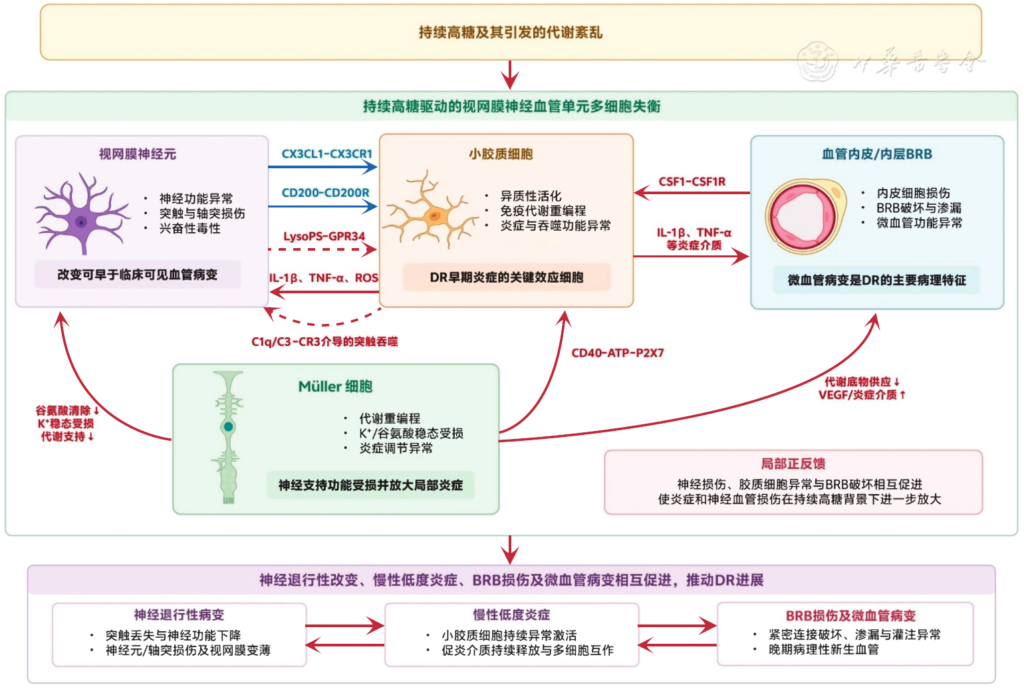
Figure 1 Mechanism of neuroimmune interaction in DR Continuous high glucose and the metabolic disorders it causes can lead to dysfunction of retinal neurons, microglia, Müller cells, vascular endothelial cells and inner layer BRB, and mediate the mutual promotion of neurodegenerative changes, chronic low-grade inflammation, BRB damage and microangiopathy through multiple intercellular signals. Blue solid arrows indicate the role of maintaining homeostasis or inhibiting inflammation; red solid arrows indicate the role of promoting inflammation or tissue damage; red dotted arrows indicate that there is currently limited direct causal evidence for the relevant mechanism in early DR.
DR: diabetic retinopathy;BRB: Blood-retinal barrier; CX3CL1: C-X3-C motif chemokine ligand 1; CX3CR1: C-X3-C motif chemokine receptor 1; CD200: Cluster of Differentiation 200; CD200R: CD200 receptor; LysoPS: Lysophosphatidylserine; GPR34: G protein-coupled receptor 34; IL-1β: interleukin-1β; TNF-α: Tumor necrosis factor alpha; ROS: Reactive oxygen species; C1q: Complement component 1q; C3: Complement component 3; CR3: Complement receptor 3; CSF1: Colony stimulating factor 1; CSF1R: Colony stimulating factor 1 receptor; CD40: differentiation antigen cluster 40; ATP: Adenosine triphosphate; P2X7: P2X purinergic receptor 7; VEGF: vascular endothelial growth factor
References click to collapse
[1] Jampol LM, Glassman AR, Sun J. Evaluation and care of patients with diabetic retinopathy[J]. N Engl J Med, 2020, 382(17):1629-1637. DOI: 10.1056/NEJMra1909637.
[2] Hou X, Wang L, Zhu D, et al. Prevalence of diabetic retinopathy and vision-threatening diabetic retinopathy in adults with diabetes in China[J]. Nat Commun, 2023, 14(1):4296. DOI: 10.1038/s41467-023-39864-w.
[3] Wong TY, Cheung CM, Larsen M, et al. Diabetic retinopathy[J]. Nat Rev Dis Primers, 2016, 2:16012. DOI: 10.1038/nrdp.2016.12.
[4] Clements JN, Mesawich J, Twisdale C. Current and emerging pharmacologic therapies for diabetic retinopathy[J/OL]. Pharmacotherapy, 2026, 46(3):e70119[2026-04-10]. https://pubmed.ncbi.nlm.nih.gov/41693076/. DOI: 10.1002/phar.70119.
[5] Fu DJ, Mishra AV, Quek C, et al. Visual and anatomical failure of anti-VEGF therapy for retinal vascular diseases: a survival analysis of real-world data[J]. Eye (Lond), 2025, 39(5):977-985. DOI: 10.1038/s41433-024-03529-9.
[6] Callan A, Heckman J, Tah G, et al. VEGF in diabetic retinopathy and age-related macular degeneration[J]. Int J Mol Sci, 2025, 26(11):4992. DOI: 10.3390/ijms26114992.
[7] Antonetti DA, Silva PS, Stitt AW. Current understanding of the molecular and cellular pathology of diabetic retinopathy[J]. Nat Rev Endocrinol, 2021, 17(4):195-206. DOI: 10.1038/s41574-020-00451-4.
[8] Sohn EH, van Dijk HW, Jiao C, et al. Retinal neurodegeneration may precede microvascular changes characteristic of diabetic retinopathy in diabetes mellitus[J/OL]. Proc Natl Acad Sci U S A, 2016, 113(19): E2655-E2664[2026-04-12]. https://pubmed.ncbi.nlm.nih.gov/27114552/ DOI: 10.1073/pnas.1522014113.
[9] Qian HY, Wei XH, Huang JO. Inflammatory mechanisms in diabetic retinopathy: pathogenic roles and therapeutic perspectives[J]. Am J Transl Res, 2025, 17(8):6262-6274. DOI: 10.62347/GBFO5856.
[10] Nian S, Lo A, Mi Y, et al. Neurovascular unit in diabetic retinopathy: pathophysiological roles and potential therapeutical targets[J]. Eye Vis (Lond), 2021, 8(1):15. DOI: 10.1186/s40662-021-00239-1.
[11] Chen BR, Wu T, Chen TH, et al. Neuroimmune interactions and their roles in neurodegenerative diseases[J]. Fundam Res, 2024, 4(2): 251-261. DOI: 10.1016/j.fmre.2023.04.002.
[12] Huh JR, Veiga-Fernandes H. Neuroimmune circuits in inter-organ communication[J]. Nat Rev Immunol, 2020, 20(4):217-228. DOI: 10.1038/s41577-019-0247-z.
[13] Ptito M, Bleau M, Bouskila J. The retina: a window into the brain[J]. Cells, 2021, 10(12):3269. DOI: 10.3390/cells10123269.
[14] Wu S, Mo X. Optic nerve regeneration in diabetic retinopathy: potentials and challenges ahead[J]. Int J Mol Sci, 2023, 24(2):1447. DOI: 10.3390/ijms24021447.
[15] Jeon J, Park YS, Kim SH, et al. Deciphering perivascular macrophages and microglia in the retinal ganglion cell layers[J]. Front Cell Dev Biol, 2024, 12:1368021. DOI: 10.3389/fcell.2024.1368021.
[16] Zhang Y, Zhao L, Wang X, et al. Repopulating retinal microglia restore endogenous organization and function under CX3CL1-CX3CR1 regulation[J/OL]. Sci Adv, 2018, 4(3):eaap8492[2026-04-16]. https://pubmed.ncbi.nlm.nih.gov/29750189/. DOI: 10.1126/sciadv.aap8492.
[17] Chen Y, Coorey NJ, Zhang M, et al. Metabolism dysregulation in retinal diseases and related therapies[J]. Antioxidants (Basel), 2022, 11(5): 942. DOI: 10.3390/antiox11050942.
[18] Calbiague García V, Chen Y, Cádiz B, et al. Imaging of lactate metabolism in retinal Müller cells with a FRET nanosensor[J]. Exp Eye Res, 2023, 226:109352. DOI: 10.1016/j.exer.2022.109352.
[19] Lindsay KJ, Du J, Sloat SR, et al. Pyruvate kinase and aspartate-glutamate carrier distributions reveal key metabolic links between neurons and glia in retina[J]. Proc Natl Acad Sci U S A, 2014, 111(43):15579-15584. DOI: 10.1073/pnas.1412441111.
[20] Beverley KM, Pattnaik BR. Inward rectifier potassium (Kir) channels in the retina: living our vision[J]. Am J Physiol Cell Physiol, 2022, 323(3): C772-C782. DOI: 10.1152/ajpcell.00112.2022.
[21] Katoozi S, Rao SB, Skauli N, et al. Functional specialization of retinal Müller cell endfeet depends on an interplay between two syntrophin isoforms[J]. Mol Brain, 2020, 13(1):40. DOI: 10.1186/s13041-020-00581-w.
[22] Subair TI, Oyetayo B, Morales-Ramírez N, et al. Glutamate uptake activity in retina Müller cells: circadian modulation[J]. Exp Eye Res, 2025, 260:110603. DOI: 10.1016/j.exer.2025.110603.
[23] Grimes WN, Berson DM, Sabnis A, et al. Layer-specific anatomical and physiological features of the retina’s neurovascular unit[J]. Curr Biol, 2025, 35(1):109-120. DOI: 10.1016/j.cub.2024.11.023.
[24] Xiao R, Huang X, Gao S, et al. Microglia in retinal diseases: from pathogenesis towards therapeutic strategies[J]. Biochem Pharmacol, 2024, 230(Pt 1):116550. DOI: 10.1016/j.bcp.2024.116550.
[25] Fan Q, Li Z. Breach and restoration of retinal immune privilege: barrier failure, innate dysregulation, and adaptive autoimmunity[J]. Front Immunol, 2025, 16:1703382. DOI: 10.3389/fimmu.2025.1703382.
[26] Lu T, Shang J, Pu S, et al. The role of microglia in the development of diabetic retinopathy and its potential clinical application[J]. Hum Cell, 2025, 38(4):101. DOI: 10.1007/s13577-025-01226-7.
[27] Silverman SM, Wong WT. Microglia in the retina: roles in development, maturity, and disease[J]. Annu Rev Vis Sci, 2018, 4:45-77. DOI: 10.1146/annurev-vision-091517-034425.
[28] Murenu E, Gerhardt MJ, Biel M, et al. More than meets the eye: the role of microglia in healthy and diseased retina[J]. Front Immunol, 2022, 13: 1006897. DOI: 10.3389/fimmu.2022.1006897.
[29] Jobling AI, Greferath U, Dixon MA, et al. Microglial regulation of the retinal vasculature in health and during the pathology associated with diabetes[J]. Prog Retin Eye Res, 2025, 106:101349. DOI: 10.1016/j.preteyeres.2025.101349.
[30] Ramírez AI, de Hoz R, Fernández-Albarral JA, et al. Time course of bilateral microglial activation in a mouse model of laser-induced glaucoma[J]. Sci Rep, 2020, 10(1):4890. DOI: 10.1038/s41598-020-61848-9.
[31] Balzamino BO, Cacciamani A, Dinice L, et al. Retinal inflammation and reactive müller cells: neurotrophins’ release and neuroprotective strategies[J]. Biology (Basel), 2024, 13(12):1030. DOI: 10.3390/biology 13121030.
[32] Apostolidi A, Stergiopoulos G, Bellou S, et al. Advances in modeling the inner blood-retinal barrier: from static tissue cell cultures to microphysiological systems[J]. Pharmaceuticals (Basel), 2025, 18(9): 1374. DOI: 10.3390/ph18091374.
[33] Kaur G, Sharma D, Bisen S, et al. Vascular cell-adhesion molecule 1 (VCAM-1) regulates JunB-mediated IL-8/CXCL1 expression and pathological neovascularization[J]. Commun Biol, 2023, 6(1):516. DOI: 10.1038/s42003-023-04905-z.
[34] D’Esposito F, Cappellani F, Visalli F, et al. Pericytes as key players in retinal diseases: a comprehensive narrative review[J]. Biology (Basel), 2025, 14(7):736. DOI: 10.3390/biology14070736.
[35] Sterling JK, Rajesh A, Droho S, et al. Retinal perivascular macrophages regulate immune cell infiltration during neuroinflammation in mouse models of ocular disease[J/OL]. J Clin Invest, 2024, 134(20):e180904 [2026-04-16]. https://pubmed.ncbi.nlm.nih.gov/39207852/. DOI: 10.1172/JCI180904.
[36] Quiriconi P, Hristov V, Aburaya M, et al. The role of microglia in the development of diabetic retinopathy[J]. NPJ Metab Health Dis, 2024, 2(1):7. DOI: 10.1038/s44324-024-00009-2.
[37] Wang Y, Yang X, Zhang Y, et al. Single-cell RNA sequencing reveals roles of unique retinal microglia types in early diabetic retinopathy[J]. Diabetol Metab Syndr, 2024, 16(1):49. DOI: 10.1186/s13098-024-01282-3.
[38] Lv K, Ying H, Hu G, et al. Integrated multi-omics reveals the activated retinal microglia with intracellular metabolic reprogramming contributes to inflammation in STZ-induced early diabetic retinopathy[J]. Front Immunol, 2022, 13:942768. DOI: 10.3389/fimmu.2022.942768.
[39] Bai Y, Wang X, Qi F, et al. Mechanisms of action of retinal microglia in diabetic retinopathy (review)[J]. Int J Mol Med, 2025, 56(6):202. DOI: 10.3892/ijmm.2025.5643.
[40] Ben S, Ma Y, Bai Y, et al. Microglia-endothelial cross-talk regulates diabetes-induced retinal vascular dysfunction through remodeling inflammatory microenvironment[J]. iScience, 2024, 27(3):109145. DOI: 10.1016/j.isci.2024.109145.
[41] Cai L, Xia M, Zhang F. Redox regulation of immunometabolism in microglia underpinning diabetic retinopathy[J]. Antioxidants (Basel), 2024, 13(4):423. DOI: 10.3390/antiox13040423.
[42] Hong S, Beja-Glasser VF, Nfonoyim BM, et al. Complement and microglia mediate early synapse loss in Alzheimer mouse models[J]. Science, 2016, 352(6286):712-716. DOI: 10.1126/science.aad8373.
[43] Wilton DK, Mastro K, Heller MD, et al. Microglia and complement mediate early corticostriatal synapse loss and cognitive dysfunction in Huntington’s disease[J]. Nat Med, 2023, 29(11):2866-2884. DOI: 10.1038/s41591-023-02566-3.
[44] Church KA, Rodriguez D, Vanegas D, et al. Models of microglia depletion and replenishment elicit protective effects to alleviate vascular and neuronal damage in the diabetic murine retina[J]. J Neuroinflammation, 2022, 19(1):300. DOI: 10.1186/s12974-022-02659-9.
[45] Yang S, Qi S, Wang C. The role of retinal Müller cells in diabetic retinopathy and related therapeutic advances[J]. Front Cell Dev Biol, 2022, 10:1047487. DOI: 10.3389/fcell.2022.1047487.
[46] Corano Scheri K, Hsieh YW, Tedeschi T, et al. Müller cell glutamine metabolism links photoreceptor and endothelial injury in diabetic retinopathy[J/OL]. Life Sci Alliance, 2026, 9(2):e202503434[2026-04-16]. https://pubmed.ncbi.nlm.nih.gov/41266111/. DOI: 10.26508/lsa.202503434.
[47] Li X, Lv J, Li J, et al. Kir4.1 may represent a novel therapeutic target for diabetic retinopathy (review)[J]. Exp Ther Med, 2021, 22(3):1021. DOI: 10.3892/etm.2021.10453.
[48] Augustine J, Pavlou S, Harkin K, et al. IL-33 regulates Müller cell-mediated retinal inflammation and neurodegeneration in diabetic retinopathy[J]. Dis Model Mech, 2023, 16(9):dmm050174. DOI: 10.1242/dmm.050174.
[49] Rezzola S, Guerra J, Krishna Chandran AM, et al. VEGF-independent activation of müller cells by the vitreous from proliferative diabetic retinopathy patients[J]. Int J Mol Sci, 2021, 22(4):2179. DOI: 10.3390/ijms22042179.
[50] Portillo JC, Lopez Corcino Y, Miao Y, et al. CD40 in retinal müller cells induces P2X7-dependent cytokine expression in macrophages/microglia in diabetic mice and development of early experimental diabetic retinopathy[J]. Diabetes, 2017, 66(2):483-493. DOI: 10.2337/db16-0051.
[51] Huang Z, Liang J, Chen S, et al. RIP3-mediated microglial necroptosis promotes neuroinflammation and neurodegeneration in the early stagesof diabetic retinopathy[J]. Cell Death Dis, 2023, 14(3):227. DOI: 10.1038/s41419-023-05660-z.
[52] Kong H, Zhao H, Chen T, et al. Targeted P2X7/NLRP3 signaling pathway against inflammation, apoptosis, and pyroptosis of retinal endothelial cells in diabetic retinopathy[J]. Cell Death Dis, 2022, 13(4): 336. DOI: 10.1038/s41419-022-04786-w.
[53] Tang Z, Yang D, Nguyen TX, et al. Relationship of OCT-based diabetic retinal neurodegeneration to the development and progression of diabetic retinopathy: a cohort study[J]. Invest Ophthalmol Vis Sci, 2025, 66(2):32. DOI: 10.1167/iovs.66.2.32.
[54] Zhong X, Li H, Tan S, et al. Initial retinal nerve fiber layer loss and risk of diabetic retinopathy over a four-year period[J]. Invest Ophthalmol Vis Sci, 2024, 65(12):5. DOI: 10.1167/iovs.65.12.5.
[55] Li RR, Yang Y, Zhang MG, et al. Abnormalities of retinal function in type 2 diabetes mellitus patients without clinical diabetic retinopathy detected by multifocal electroretinogram[J]. BMC Ophthalmol, 2024, 24(1):71. DOI: 10.1186/s12886-024-03335-7.
[56] Arias-Alvarez M, Sopeña-Pinilla M, Fernandez-Espinosa G, et al. Retinal function in long-term type 1 diabetes without retinopathy: insights from pattern electroretinogram and pattern visual evoked potentials assessments[J]. Diagnostics (Basel), 2024, 14(5):492. DOI: 10.3390/diagnostics14050492.
[57] Vinković M, Kopić A, Benašić T, et al. HD-OCT angiography and SD-OCT in patients with mild or no clinically apparent diabetic retinopathy[J]. Biomedicines, 2025, 13(5): 1251. DOI: 10.3390/biomedicines13051251.
[58] Paolicelli RC, Bisht K, Tremblay MÈ. Fractalkine regulation of microglial physiology and consequences on the brain and behavior[J]. Front Cell Neurosci, 2014, 8:129. DOI: 10.3389/fncel.2014.00129.
[59] Rodriguez D, Church KA, Pietramale AN, et al. Fractalkine isoforms differentially regulate microglia-mediated inflammation and enhance visual function in the diabetic retina[J]. J Neuroinflammation, 2024, 21(1):42. DOI: 10.1186/s12974-023-02983-8.
[60] Pfeifer CW, Walsh JT, Santeford A, et al. Dysregulated CD200-CD200R signaling in early diabetes modulates microglia-mediated retinopathy[J/OL]. Proc Natl Acad Sci U S A, 2023, 120(45):e2308214120[2026-04-18]. https://pubmed.ncbi.nlm.nih.gov/37903272/. DOI: 10.1073/pnas.2308214120.
[61] Kotwica-Mojzych K, Jodłowska-Jędrych B, Mojzych M. CD200:CD200R interactions and their importance in immunoregulation[J]. Int J Mol Sci, 2021, 22(4):1602. DOI: 10.3390/ijms22041602.
[62] Broderick C, Hoek RM, Forrester JV, et al. Constitutive retinal CD200 expression regulates resident microglia and activation state of inflammatory cells during experimental autoimmune uveoretinitis[J]. Am J Pathol, 2002, 161(5):1669-1677. DOI: 10.1016/S0002-9440(10) 64444-6.
[63] Chen L, Zhang H, Zhang Y, et al. Ganglion cell-derived LysoPS induces retinal neovascularisation by activating the microglial GPR34-PI3K-AKT-NINJ1 axis[J]. J Neuroinflammation, 2024, 21(1): 278. DOI: 10.1186/s12974-024-03265-7.
[64] Massey SC, Redburn DA. Transmitter circuits in the vertebrate retina[J]. Prog Neurobiol, 1987, 28(1):55-96. DOI: 10.1016/0301-0082(87) 90005-0.
[65] Bi Q, Cheng G, Chen N, et al. Microglial neurotransmitter receptors in health and disease[J]. Physiology (Bethesda), 2026, 41(5):0. DOI: 10.1152/physiol.00030.2025.
[66] Fontainhas AM, Wang M, Liang KJ, et al. Microglial morphology and dynamic behavior is regulated by ionotropic glutamatergic and GABAergic neurotransmission[J/OL]. PLoS One, 2011, 6(1):e15973 [2026-04-18]. https://pubmed.ncbi.nlm.nih.gov/21283568/. DOI: 10.1371/journal.pone.0015973.
[67] Hajari JN, Ilginis T, Pedersen TT, et al. Novel blood-biomarkers to detect retinal neurodegeneration and inflammation in diabetic retinopathy[J]. Int J Mol Sci, 2025, 26(6): 2625. DOI: 10.3390/ijms26062625.
[68] Gaetani L, Blennow K, Calabresi P, et al. Neurofilament light chain as a biomarker in neurological disorders[J]. J Neurol Neurosurg Psychiatry, 2019, 90(8):870-881. DOI: 10.1136/jnnp-2018-320106.
[69] Hernández C, Simó-Servat O, Porta M, et al. Serum glial fibrillary acidic protein and neurofilament light chain as biomarkers of retinal neurodysfunction in early diabetic retinopathy: results of the EUROCONDOR study[J]. Acta Diabetol, 2023, 60(6):837-844. DOI: 10.1007/s00592-023-02076-1.
[70] Kuhle J, Kropshofer H, Haering DA, et al. Blood neurofilament light chain as a biomarker of MS disease activity and treatment response[J/OL]. Neurology, 2019, 92(10):e1007-e1015[2026-04-18]. https://pubmed.ncbi.nlm.nih.gov/30737333/. DOI: 10.1212/WNL.0000000000007032.
[71] Buonocore J, Fratto E, Arcuri F, et al. Plasma NfL and GFAP in the preclinical stages of neurodegenerative diseases: insights from the UK Biobank[J]. J Neurol, 2025, 272(12):755. DOI: 10.1007/s00415-025-13498-y.
[72] Wang W, Sun G, Xu A, et al. Proliferative diabetic retinopathy and diabetic macular edema are two factors that increase macrophage-like cell density characterized by en face optical coherence tomography[J]. BMC Ophthalmol, 2023, 23(1):46. DOI: 10.1186/s12886-023-02794-8.
[73] Zhang NT, Nesper PL, Ong JX, et al. Macrophage-like cells are increased in patients with vision-threatening diabetic retinopathy and correlate with macular edema[J]. Diagnostics (Basel), 2022, 12(11): 2793. DOI: 10.3390/diagnostics12112793.
[74] Mat Nor MN, Green CR, Squirrell D, et al. Retinal hyperreflective foci are biomarkers of ocular disease: a scoping review with evidence from humans and insights from animal models[J]. J Ophthalmol, 2025, 2025: 9573587. DOI: 10.1155/joph/9573587.
[75] Lachin JM, Nathan DM, DCCT/EDIC research group. Understanding metabolic memory: the prolonged influence of glycemia during the diabetes control and complications trial (DCCT) on future risks of complications during the study of the epidemiology of diabetes interventions and complications (EDIC)[J]. Diabetes Care, 2021,44(10): 2216-2224. DOI: 10.2337/dc20-3097.
[76] Wang J, Yin Z, Yang J, et al. BCAT1 activation reprograms branched-chain amino acid metabolism and epigenetically promotes inflammation in diabetic retinopathy[J]. Invest Ophthalmol Vis Sci, 2025, 66(6):59. DOI: 10.1167/iovs.66.6.59.
[77] Jiang M, Xie H, Zhang C, et al. Enhancing fractalkine/CX3CR1 signalling pathway can reduce neuroinflammation by attenuating microglia activation in experimental diabetic retinopathy[J]. J Cell Mol Med, 2022, 26(4):1229-1244. DOI: 10.1111/jcmm.17179.
[78] Yilmaz O, Erdogan M, Algemi M, et al. Relationship between the degree of diabetic retinopathy and serum fractalkine (CX3CL1) in patients with type 2 diabetes: a single-center cross-sectional study[J]. Medicina (Kaunas), 2026, 62(2):312. DOI: 10.3390/medicina62020312.
[79] Fang W, Liu S, Wu Y, et al. IL-12 ameliorates diabetic retinal neurodegeneration by activating microglial phagocytosis via the TREM2/ DAP12 pathway[J]. Mol Neurobiol, 2025, 63(1):215. DOI: 10.1007/s12035-025-05512-1.
[80] Li HY, Wang J, Xiao T, et al. STING immune activation of microglia aggravating neurovascular unit damage in diabetic retinopathy[J]. Free Radic Biol Med, 2025, 233:86-101. DOI: 10.1016/j.freeradbiomed.2025.03.042.
[81] Fan Y, Ge P, Wang X, et al. Microglia-specific interleukin-4 delivery by engineered extracellular vesicles restores inner blood-retinal barrier in diabetic retinopathy via GAS6-MERTK pathway[J]. J Nanobiotechnology, 2025, 24(1):101. DOI: 10.1186/s12951-025-03976-w.