Authors:Liu Wenhui, Gao Jingzhao, Su Lin, Li Xiaorong
Corresponding author: Li Xiaorong , Email: xiaorli@163.com
Published:2026-09-10
DOI:10.3760/cma.j.cn115989-20260331-00151
ABSTRACT
Oxygen-induced retinopathy (OIR) is an important animal model for retinal neovascular diseases. In recent years, increasing evidence has shown that alterations in the retinal immune microenvironment are involved throughout the development and progression of OIR, among which myeloid immune cells, such as microglia and macrophages, serve as key regulators linking inflammatory responses, vascular remodeling, and tissue repair. Myeloid cells in OIR exhibit marked heterogeneity in origin and functional plasticity: resident microglia participate in inflammatory responses through local proliferation and activation, whereas peripheral monocytes can be recruited into the retina under ischemic conditions and differentiate into macrophages. Current studies have shown that these cells are jointly regulated by glycolysis, lactate metabolism, succinate signaling, and inflammatory pathways, and may display predominantly pro-inflammatory or reparative functional states at different stages, thereby exerting a distinct dual role. On the one hand, they can amplify inflammatory responses, aggravate vascular obliteration, and promote pathological retinal neovascularization; on the other hand, they can contribute to vascular homeostasis, physiological revascularization, and tissue repair. Therefore, precise targeting of myeloid immune cells has gradually emerged as a new research focus. This review summarizes the origins, functional state transitions, metabolic-inflammatory regulatory networks, and dual roles of microglia and macrophages in OIR, and further outlines recent advances in related therapeutic strategies, with the aim of providing references for mechanistic studies and precision treatment of retinal neovascular diseases.
KEYWORDS:
Oxygen-induced retinopathy;Microglia;Macrophages;Retinal neovascularization;Immune microenvironment
COPYRIGHTS:
Copyright by Chinese Medical Association
No content published by the journals of Chinese Medical Association may be reproduced or abridged without authorization. Please do not use or copy the layout and design of the journals without permission.
All articles published represent the opinions of the authors, and do not reflect the official policy of the Chinese Medical Association or the Editorial Board, unless this is clearly specified.
Authors Info & Affiliations
Liu Wenhui
Tianjin Key Laboratory of Retinal Functions and Diseases, Tianjin Branch of National Clinical Research Center for Ocular Disease, Eye Institute and School of Optometry, Tianjin Medical University Eye Hospital, Tianjin 300384, China
Gao Jingzhao
Tianjin Key Laboratory of Retinal Functions and Diseases, Tianjin Branch of National Clinical Research Center for Ocular Disease, Eye Institute and School of Optometry, Tianjin Medical University Eye Hospital, Tianjin 300384, China
Su Lin
Tianjin Key Laboratory of Retinal Functions and Diseases, Tianjin Branch of National Clinical Research Center for Ocular Disease, Eye Institute and School of Optometry, Tianjin Medical University Eye Hospital, Tianjin 300384, China
Li Xiaorong
Tianjin Key Laboratory of Retinal Functions and Diseases, Tianjin Branch of National Clinical Research Center for Ocular Disease, Eye Institute and School of Optometry, Tianjin Medical University Eye Hospital, Tianjin 300384, China
Figures & Tables
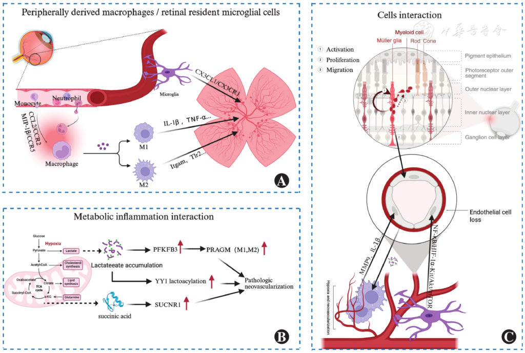
Figure 1 Role and mechanism of macrophages and microglia in OIR A: Origin and phenotype of myeloid cells and retina-resident microglia (CX3CL1/CX3CR1 axis) and peripheral monocytes infiltrate and differentiate into macrophages through the CCL2/CCR2 axis, and both present M1 or M2 functional states. B: Metabolism-inflammation regulation Under hypoxia conditions, glycolysis is enhanced. Lactate drives the transformation of PFKFB3 to PRAGM phenotype and the YY1 lactoylation pathway. Succinic acid promotes pathological neovascularization through SUCNR1 signal. C: Cell interaction network Myeloid cells interact with Müller glial cells, vascular endothelial cells and photoreceptor cells to jointly regulate vascular occlusion and neovascularization
OIR: oxygen-induced retinopathy; CCL2: C-C motif chemokine ligand 2;CCR: C-C chemokine receptor;MIP: macrophage inflammatory protein;IL: interleukin;TNF-α: tumor necrosis factor alpha; Tlr2: Toll-like receptor 2; CX3CL1: chemokine (C-X3-C motif) ligand 1; CX3CR1: CX3C chemokine receptor 1;MMP: matrix metalloproteinase;NF-κB: nuclear factor κB;HIF-1α-Kit: hypoxia-inducible factor-1 α- KIT proto-oncogene;mTOR: mammalian rapamycin target protein; PFKFB3: 6-phosphate fructose-2-kinase/fructose-2,6-bisphosphatase3;PRAGM: pathological angiogenesis associated glycolytic macrophages/microglia; YY1: transcription factor Yinyang 1; SUCNR1: Succinate receptor 1
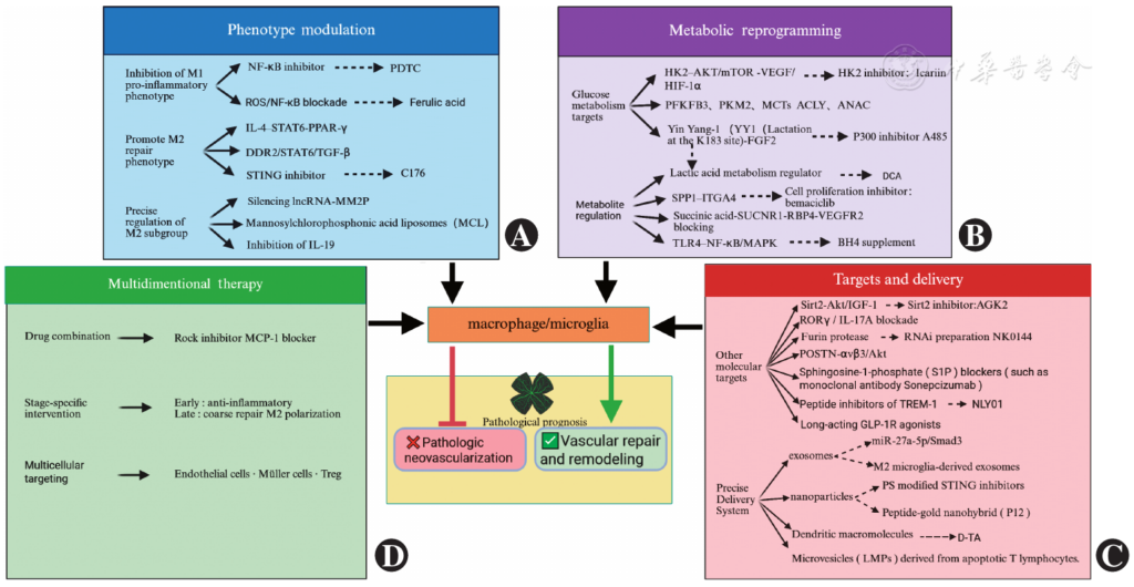
Figure 2 Therapeutic strategy targeting macrophages/microglia A: Phenotype regulation suppresses the M1 pro-inflammatory phenotype (NF-κB inhibitor PDTC and ferulic acid block the ROS/NF-κB axis), promote M2 repair phenotype, and accurately regulate M2 subgroups (Silencing lncRNA-MM2P, mannose-clodronic acid liposome MCL clears M2 phenotype and blocks IL-19) B: Metabolic reprogramming targets glycolysis (HK2 inhibitor icariin, PFKFB3/PKM2/MCTs/ACLY node), interfering with lactate signaling (DCA regulates lactate metabolism and blocks lactoylation at YY1K183 site), inhibits the SPP1-ITGA4 axis (Proliferation inhibitor abemaciclib) and succinine-SUCNR1-RBP4-VEGFR2 axis, supplemented with BH4 to block the TLR4-NF-κB/MAPK pathway C: Targeting and delivery Sirt2/Akt, RORγ/IL-17A blockade, furin protease, POSTN New targets such as (αvβ3/Akt) RNAi agent NK0144, S1P blocker sonepcizumab, TREM-1 peptide inhibitor, and long-acting GLP-1R agonist NLY01; Precision delivery systems include microglial exosomes (miR-27a-5p/Smad3), M2 exosomes, PS-modified STING inhibitor nanoparticles, peptide-gold nanohybrid P12, dendritic molecule D-TA and apoptotic T cell microvesicles LMPs D: multi-dimensional combination therapy ROCK inhibitors combined with MCP-1 blockade, phased intervention in early anti-inflammation and late repair promotion, endothelial cells-Müller cells-Tregs multicellular targeting
PDTC: pyrrolidine dithiocarbamate; ROS: Reactive oxygen species; NF-κB: Nuclear factor κB; MCL: Mannose-clodronate liposomes;IL: interleukin;STAT: signal transducer and activator of transcription;PPAR-γ: peroxisome proliferator-activated receptor γ;DDR2: discoid domain receptor 2;TGF-β: transforming growth factor beta;STING: interferon gene stimulating factor;lncRNA: long-chain non-coding RNA; MM2P: M2 macrophage polarization; HK2: hexokinase 2;AKT: protein kinase B;VEGF: vascular endothelial growth factor; PFKFB3: Fructose-6-phosphate-2-kinase/Fructose-2,6-bisphosphatase3; PKM2: Pyruvate kinase type M2;MCTs: monocarboxylic acid transporter;ACLY: ATP citrate lyase;ANAC: Acetyl-CoA and NAD+-related metabolic cycles;FGF: fibroblast growth factor;DCA: dichloroacetic acid; SPP1: secreted phosphoprotein 1; ITGA4: integrin alpha 4; SUCNR1: succinate receptor 1; RBP4: retinol binding protein 4; VEGFR2: vascular endothelial growth factor receptor 2; TLR4: Toll-like receptor 4; MAPK: Mitogen-activated protein kinase; BH4: Tetrahydrobiopterin; MCP-1: Monocyte chemoattractant protein-1;Tregs: regulatory T cells;Sirt2: silent information regulator 2;IGF-1: insulin-like growth factor 1; AGK2: specific inhibitor of Sirt2;RORγ: retinoic acid-related orphan receptor γ;POSTN: periostin; S1P: sphingosine-1-phosphate;TREM-1: myeloid cell trigger receptor 1;GLP-1R: glucagon-like peptide-1 receptor;PS: phosphatidylserine;LMPs: lymphocyte-derived microvesicles
References click to collapse
[1] Ong JX, Nesper PL, Fawzi AA, et al. Macrophage-like cell density is increased in proliferative diabetic retinopathy characterized by optical coherence tomography angiography[J]. Invest Ophthalmol Vis Sci, 2021, 62(10):2. DOI: 10.1167/iovs.62.10.2.
[2] Zhang NT, Nesper PL, Ong JX, et al. Macrophage-like cells are increased in patients with vision-threatening diabetic retinopathy and correlate with macular edema[J]. Diagnostics (Basel), 2022, 12(11): 2793. DOI: 10.3390/diagnostics12112793.
[3] Hata M, Hata M, Dejda A, et al. Corticosteroids reduce pathological angiogenesis yet compromise reparative vascular remodeling in a model of retinopathy[J/OL]. Proc Natl Acad Sci U S A, 2024, 121(52): e2411640121[2026-03-12]. https://pubmed.ncbi.nlm.nih.gov/39693344/. DOI: 10.1073/pnas.2411640121.
[4] Yamaguchi M, Nakao S, Arima M, et al. Heterotypic macrophages/microglia differentially contribute to retinal ischaemia and neovascularisation[J]. Diabetologia, 2024, 67(10):2329-2345. DOI: 10.1007/s00125-024-06215-3.
[5] Boeck M, Thien A, Wolf J, et al. Temporospatial distribution and transcriptional profile of retinal microglia in the oxygen-induced retinopathy mouse model[J]. Glia, 2020, 68(9): 1859-1873. DOI: 10.1002/glia.23810.
[6] Shao A, Jin L, Ge Y, et al. C176-loaded and phosphatidylserine-modified nanoparticles treat retinal neovascularization by promoting M2 macrophage polarization[J]. Bioact Mater, 2024, 39: 392-405. DOI: 10.1016/j.bioactmat.2024.05.038.
[7] NiB,YangZ,ZhouT,etal.Therapeutic intervention in neuroinflammation for neovascular ocular diseases through targeting the cGAS-STING necroptosis pathway[J]. J Neuroinflammation, 2024, 21(1):164. DOI: 10.1186/s12974-024-03155-y.
[8] Lorenc VE, Lima E Silva R, Hackett SF, et al. Hepatocyte growth factor is upregulated in ischemic retina and contributes to retinal vascular leakage and neovascularization[J]. FASEB Bioadv, 2020, 2(4): 219-233. DOI: 10.1096/fba.2019-00074.
[9] Wang JH, Kumar S, Liu GS. Bulk gene expression deconvolution reveals infiltration of M2 macrophages in retinal neovascularization[J]. Invest Ophthalmol Vis Sci, 2021, 62(14): 22. DOI: 10.1167/iovs.62.14.22.
[10] Davies MH, Eubanks JP, Powers MR. Microglia and macrophages are increased in response to ischemia-induced retinopathy in the mouse retina[J]. Mol Vis, 2006, 12:467-477. cells
[11] Ishikawa K, Yoshida S, Nakao S, et al. Bone marrow-derived monocyte lineage recruited by MIP-1β promote physiological revascularization in mouse model of oxygen-induced retinopathy[J]. Lab Invest, 2012, 92(1):91-101. DOI: 10.1038/labinvest.2011.141.
[12] Zhao L, Ma W, Fariss RN, et al. Retinal vascular repair and neovascularization are not dependent on CX3CR1 signaling in a model of ischemic retinopathy[J]. Exp Eye Res, 2009, 88(6):1004-1013. DOI: 10.1016/j.exer.2008.12.013.
[13] Li J, Yu S, Lu X, et al. The phase changes of M1/M2 phenotype of microglia/macrophage following oxygen-induced retinopathy in mice[J]. Inflamm Res, 2021, 70(2):183-192. DOI: 10.1007/s00011-020-01427-w.
[14] Zhu Y, Zhang L, Lu Q, et al. Identification of different macrophage subpopulations with distinct activities in a mouse model of oxygen-induced retinopathy[J]. Int J Mol Med, 2017, 40(2):281-292. DOI: 10.3892/ijmm.2017.3022.
[15] Gao S, Li C, Zhu Y, et al. PEDF mediates pathological neovascularization by regulating macrophage recruitment and polarization in the mouse model of oxygen-induced retinopathy[J]. Sci Rep, 2017, 7:42846. DOI: 10.1038/srep42846.
[16] Zhou Y, Yoshida S, Nakao S, et al. M2 macrophages enhance pathological neovascularization in the mouse model of oxygen-induced retinopathy[J]. Invest Ophthalmol Vis Sci, 2015, 56(8):4767-4777. DOI: 10.1167/iovs.14-16012.
[17] Zou J, Tan W, Li B, et al. Interleukin-19 promotes retinal neovascularization in a mouse model of oxygen-induced retinopathy[J]. Invest Ophthalmol Vis Sci, 2022, 63(8):9. DOI: 10.1167/iovs.63.8.9.
[18] Zhou H, Zhang L, Ding C, et al. Upregulation of HMOX1 associated with M2 macrophage infiltration and ferroptosis in proliferative diabetic retinopathy[J]. Int Immunopharmacol, 2024, 134: 112231. DOI: 10.1016/j.intimp.2024.112231.
[19] Liu Z, Shi H, Xu J, et al. Single-cell transcriptome analyses reveal microglia types associated with proliferative retinopathy[J/OL]. JCI Insight, 2022, 7(23):e160940[2026-03-12]. https://pubmed.ncbi.nlm.nih.gov/36264636/. DOI: 10.1172/jci.insight.160940.
[20] Liu Z, Xu J, Ma Q, et al. Glycolysis links reciprocal activation of myeloid cells and endothelial cells in the retinal angiogenic niche[J/OL]. Sci Transl Med, 2020, 12(555):eaay1371[2026-03-12]. https://pubmed.ncbi.nlm.nih.gov/32759274/. DOI: 10.1126/scitranslmed.aay1371.
[21] Wang X, Fan W, Li N, et al. YY1 lactylation in microglia promotes angiogenesis through transcription activation-mediated upregulation of FGF2[J]. Genome Biol, 2023, 24(1):87. DOI: 10.1186/s13059-023-02931-y.
[22] Hartman GD, Muniyandi A, Sishtla K, et al. Ref-1 redox activity regulates retinal neovascularization by modulating transcriptional activation of HIF-1α[J/OL]. FASEB J, 2025, 39(3):e70348[2026-03-12]. https://pubmed.ncbi.nlm.nih.gov/39902892/. DOI: 10.1096/fj.202401989RR.
[23] Liu J, Tsang J, Fung F, et al. Retinal microglia protect against vascular damage in a mouse model of retinopathy of prematurity[J]. Front Pharmacol, 2022, 13:945130. DOI: 10.3389/fphar.2022.945130.
[24] Zhou Z, Jing Y, Niu Y, et al. Distinguished functions of microglia in the two stages of oxygen-induced retinopathy: a novel target in the treatment of ischemic retinopathy[J]. Life (Basel), 2022, 12(10):1676. DOI: 10.3390/life12101676.
[25] Xu W, Hu Z, Lv Y, et al. Microglial density determines the appearance of pathological neovascular tufts in oxygen-induced retinopathy[J]. Cell Tissue Res, 2018, 374(1):25-38. DOI: 10.1007/s00441-018-2847-5.
[26] Zou K, Li X, Ren B, et al. Single-cell analysis identifies MKI67+ microglia as drivers of neovascularization in proliferative diabetic retinopathy[J]. J Transl Med, 2025, 23(1):310. DOI: 10.1186/s12967-025-06320-w.
[27] Luo Q, Jiang Z, Jiang J, et al. Tsp-1+ microglia attenuate retinal neovascularization by maintaining the expression of Smad3 in endothelial cells through exosomes with decreased miR-27a-5p[J]. Theranostics, 2023, 13(11):3689-3706. DOI: 10.7150/thno.84236.
[28] Brockmann C, Dege S, Crespo-Garcia S, et al. Spatial distribution of CD115+ and CD11b+ cells and their temporal activation during oxygen-induced retinopathy in mice[J]. Graefes Arch Clin Exp Ophthalmol, 2018, 256(2): 313-323. DOI: 10.1007/s00417-017-3845-0.
[29] Shen J, Xie B, Dong A, et al. In vivo immunostaining demonstrates macrophages associate with growing and regressing vessels[J]. Invest Ophthalmol Vis Sci, 2007, 48(9):4335-4341. DOI: 10.1167/iovs.07-0113.
[30] Kataoka K, Nishiguchi KM, Kaneko H, et al. The roles of vitreal macrophages and circulating leukocytes in retinal neovascularization[J]. Invest Ophthalmol Vis Sci, 2011, 52(3):1431-1438. DOI: 10.1167/iovs.10-5798.
[31] Li J, Wang JJ, Peng Q, et al. Macrophage metalloelastase (MMP-12) deficiency mitigates retinal inflammation and pathological angiogenesis in ischemic retinopathy[J/OL]. PLoS One, 2012, 7(12):e52699[2026-03-14]. https://pubmed.ncbi.nlm.nih.gov/23285156/. DOI: 10.1371/journal.pone.0052699.
[32] Gao X, Wang YS, Li XQ, et al. Macrophages promote vasculogenesis of retinal neovascularization in an oxygen-induced retinopathy model in mice[J]. Cell Tissue Res, 2016, 364(3):599-610. DOI: 10.1007/s00441-015-2353-y.
[33] Geraldo LH, Xu Y, Mouthon G, et al. Monoclonal antibodies that block Roundabout 1 and 2 signaling target pathological ocular neovascularization through myeloid cells[J/OL]. Sci Transl Med, 2024, 16(774):eadn8388[2026-03-14]. https://pubmed.ncbi.nlm.nih.gov/39565875/. DOI: 10.1126/scitranslmed.adn8388.
[34] Vähätupa M, Cordova ZM, Barker H, et al. Furin deficiency in myeloid cells leads to attenuated revascularization in a mouse-model of oxygen-induced retinopathy[J]. Exp Eye Res, 2018, 166:160-167. DOI: 10.1016/j.exer.2017.10.013.
[35] Ritter MR, Banin E, Moreno SK, et al. Myeloid progenitors differentiate into microglia and promote vascular repair in a model of ischemic retinopathy[J]. J Clin Invest, 2006, 116(12):3266-3276. DOI: 10.1172/JCI29683.
[36] Bai Q, Wang X, Yan H, et al. Microglia-derived Spp1 promotes pathological retinal neovascularization via activating endothelial Kit/ Akt/mTOR signaling[J]. J Pers Med, 2023, 13(1):146. DOI: 10.3390/jpm13010146.
[37] Nürnberg C, Kociok N, Brockmann C, et al. Myeloid cells contribute indirectly to VEGF expression upon hypoxia via activation of Müller cells[J]. Exp Eye Res, 2018, 166: 56-69. DOI: 10.1016/j.exer.2017.10.011.
[38] Powers MR, Davies MH, Eubanks JP. Increased expression of chemokine KC, an interleukin-8 homologue, in a model of oxygen-induced retinopathy[J]. Curr Eye Res, 2005, 30(4):299-307. DOI: 10.1080/02713680590923276.
[39] Deliyanti D, Talia DM, Zhu T, et al. Foxp3+ Tregs are recruited to the retina to repair pathological angiogenesis[J]. Nat Commun, 2017, 8(1): 748. DOI: 10.1038/s41467-017-00751-w.
[40] Joyal JS, Sun Y, Gantner ML, et al. Retinal lipid and glucose metabolism dictates angiogenesis through the lipid sensor Ffar1[J]. Nat Med, 2016, 22(4):439-445. DOI: 10.1038/nm.4059.
[41] Sui A, Chen X, Demetriades AM, et al. Inhibiting NF-κB signaling activation reduces retinal neovascularization by promoting a polarization shift in macrophages[J]. Invest Ophthalmol Vis Sci, 2020, 61(6):4. DOI: 10.1167/iovs.61.6.4.
[42] Sun X, Ma L, Li X, et al. Ferulic acid alleviates retinal neovascularization by modulating microglia/macrophage polarization through the ROS/NF-κB axis[J]. Front Immunol, 2022, 13:976729. DOI: 10.3389/fimmu.2022.976729.
[43] Dai Q, Su W, Zhou Z, et al. DDR2 alleviates retinal vaso-obliteration and pathological neovascularization by modulating microglia M1/M2 phenotypic polarization in a mouse model of proliferative retinopathy[J]. Biochim Biophys Acta Mol Basis Dis, 2025, 1871(5):167787. DOI: 10.1016/j.bbadis.2025.167787.
[44] Gao S, Li N, Wang Y, et al. Inhibition of vascular endothelial growth factor alleviates neovascular retinopathy with regulated neurotrophic/ proinflammatory cytokines through the modulation of DBI-TSPO signaling[J/OL]. FASEB J, 2022, 36(7):e22367[2026-03-16]. https://pubmed.ncbi.nlm.nih.gov/35639422/. DOI: 10.1096/fj.202101294RRR.
[45] Wang Z, Tan W, Li B, et al. LncRNA-MM2P regulates retinal neovascularization through M2 macrophage polarization[J]. Exp Eye Res, 2024, 248:110072. DOI: 10.1016/j.exer.2024.110072.
[46] Li X, Wang G, Li N, et al. Icariin alleviates oxygen-induced retinopathy by targeting microglia hexokinase 2[J]. Immunology, 2024, 173(1): 141-151. DOI: 10.1111/imm.13818.
[47] Shen T, Lin R, Hu C, et al. Succinate-induced macrophage polarization and RBP4 secretion promote vascular sprouting in ocular neovascularization[J]. J Neuroinflammation, 2023, 20(1): 308. DOI: 10.1186/s12974-023-02998-1.
[48] Edgar KS, Cunning C, Gardiner TA, et al. BH4 supplementation reduces retinal cell death in ischaemic retinopathy[J]. Sci Rep, 2023, 13(1):21292. DOI: 10.1038/s41598-023-48167-5.
[49] Wang Y, Gao S, Gao S, et al. Blocking the interaction between interleukin-17A and endoplasmic reticulum stress in macrophage attenuates retinal neovascularization in oxygen-induced retinopathy[J]. Cell Biosci, 2021, 11(1):82. DOI: 10.1186/s13578-021-00593-6.
[50] Hu Y, Wei T, Gao S, et al. Anti-angiogenic and anti-inflammatory effects of CD200-CD200R1 axis in oxygen-induced retinopathy mice model[J]. Inflamm Res, 2019, 68(11):945-955. DOI: 10.1007/s00011-019-01276-2.
[51] Jerome JR, Deliyanti D, Suphapimol V, et al. Finerenone, a non-steroidal mineralocorticoid receptor antagonist, reduces vascular injury and increases regulatory T-cells: studies in rodents with diabetic and neovascular retinopathy[J]. Int J Mol Sci, 2023, 24(3):2334. DOI: 10.3390/ijms24032334.
[52] Kielczewski JL, Hu P, Shaw LC, et al. Novel protective properties of IGFBP-3 result in enhanced pericyte ensheathment, reduced microglial activation, increased microglial apoptosis, and neuronal protection after ischemic retinal injury[J]. Am J Pathol, 2011, 178(4):1517-1528. DOI: 10.1016/j.ajpath.2010.12.031.
[53] Hu Z, Xu W, Yang X, et al. SIRT2 inhibition attenuates the vasculopathy and vision impairment via Akt signaling in retinopathy of prematurity[J]. Exp Eye Res, 2023, 233:109547. DOI: 10.1016/j.exer.2023.109547.
[54] Michan S, Juan AM, Hurst CG, et al. Sirtuin1 over-expression does not impact retinal vascular and neuronal degeneration in a mouse model of oxygen-induced retinopathy[J/OL]. PLoS One, 2014, 9(1): e85031 [2026-03-16]. https://pubmed.ncbi.nlm.nih.gov/24416337/. DOI: 10.1371/journal.pone.0085031.
[55] Zhou L, Xu Z, Oh Y, et al. Myeloid cell modulation by a GLP-1 receptor agonist regulates retinal angiogenesis in ischemic retinopathy[J/OL]. JCI Insight, 2021, 6(23):e93382[2026-03-16]. https://pubmed.ncbi.nlm.nih.gov/34673570/. DOI: 10.1172/jci.insight.93382.
[56] Talia DM, Deliyanti D, Agrotis A, et al. Inhibition of the nuclear receptor RORγ and interleukin-17A suppresses neovascular retinopathy: involvement of immunocompetent microglia[J]. Arterioscler Thromb Vasc Biol, 2016, 36(6):1186-1196. DOI: 10.1161/ATVBAHA.115.307080.
[57] Sui A, Chen X, Yao Y, et al. The IL-23/IL-17 axis promotes the formation of retinal neovascularization by activating the NLRP3 inflammasome in macrophages in an experimental retinopathy mouse model[J]. Immunology, 2021, 164(4):803-816. DOI: 10.1111/imm.13402.
[58] Nakama T, Yoshida S, Ishikawa K, et al. Therapeutic effect of novel single-stranded RNAi agent targeting periostin in eyes with retinal neovascularization[J]. Mol Ther Nucleic Acids, 2017, 6:279-289. DOI: 10.1016/j.omtn.2017.01.004.
[59] Rojas MA, Shen ZT, Caldwell RB, et al. Blockade of TREM-1 prevents vitreoretinal neovascularization in mice with oxygen-induced retinopathy[J]. Biochim Biophys Acta Mol Basis Dis, 2018, 1864(9 Pt B): 2761-2768. DOI: 10.1016/j.bbadis.2018.05.001.
[60] Xie B, Shen J, Dong A, et al. Blockade of sphingosine-1-phosphate reduces macrophage influx and retinal and choroidal neovascularization[J]. J Cell Physiol, 2009, 218(1):192-198. DOI: 10.1002/jcp.21588.
[61] Wang X, Xu C, Bian C, et al. M2 microglia-derived exosomes promote vascular remodeling in diabetic retinopathy[J]. J Nanobiotechnology, 2024, 22(1):56. DOI: 10.1186/s12951-024-02330-w.
[62] Cai C, Tahiri H, Fortin C, et al. Lymphocytic microparticles suppress retinal angiogenesis via targeting Müller cells in the ischemic retinopathy mouse model[J]. Exp Cell Res, 2021, 399(2):112470. DOI: 10.1016/j.yexcr.2021.112470.
[63] Cho H, Kambhampati SP, Lai MJ, et al. Dendrimer-triamcinolone acetonide reduces neuroinflammation, pathological angiogenesis, and neuroretinal dysfunction in ischemic retinopathy[J]. Adv Ther (Weinh), 2021, 4(2):2000181. DOI: 10.1002/adtp.202000181.
[64] Du M, Zhao X, Guo M, et al. Drugless peptide-based nanohybrids alleviate diabetic retinopathy by suppressing microglial activation and endothelial inflammation[J]. Theranostics, 2025, 15(9):3943-3960. DOI: 10.7150/thno.102775.
[65] Deliyanti D, Miller AG, Tan G, et al. Neovascularization is attenuated with aldosterone synthase inhibition in rats with retinopathy[J]. Hypertension, 2012, 59(3):607-613. DOI: 10.1161/HYPERTENSIONAHA.111.188136.