Authors:Liu Tong, Lin Xuemei, Wu Songdi
Corresponding author: Wu Songdi, Email:wusongdi@gmail.com
Published:2026-10-10
DOI:10.3760/cma.j.cn115989-20240428-00118
ABSTRACT
Tear fluid, a complex mixture of various components, is intimately linked to both ocular and systemic health through changes in its composition and properties. Due to its ease of collection, minimal invasiveness, and cost-effectiveness, tear fluid is increasingly recognized as a valuable source for biomarker research. However, the absence of standardized procedures for tear fluid collection compromises the accuracy and reproducibility of research findings, thus constraining the scope and depth of its application in biomarker studies. This review explores commonly employed methods for tear collection, such as microcapillary tubes, Schirmer strips, and the phenol red thread tear test. It offers a comparison of these techniques grounded in existing research, aiming to provide insights and recommendations for choosing the most appropriate tear collection methods.
KEYWORDS:
Tear fluid;Collection method;Tear fluid composition;Storage of tear fluid;Classification of tear fluid
COPYRIGHTS:
Copyright by Chinese Medical Association
No content published by the journals of Chinese Medical Association may be reproduced or abridged without authorization. Please do not use or copy the layout and design of the journals without permission.
All articles published represent the opinions of the authors, and do not reflect the official policy of the Chinese Medical Association or the Editorial Board, unless this is clearly specified.
Authors Info & Affiliations
Liu Tong
Department of Neuro-ophthalmology, The First Affiliated Hospital of Northwest University (Xi’an No.1 Hospital), Xi’an 710002, China
Lin Xuemei
Department of Neuro-ophthalmology, The First Affiliated Hospital of Northwest University (Xi’an No.1 Hospital), Xi’an 710002, China
Wu Songdi
Department of Neuro-ophthalmology, The First Affiliated Hospital of Northwest University (Xi’an No.1 Hospital), Xi’an 710002, China
Figures & Tables
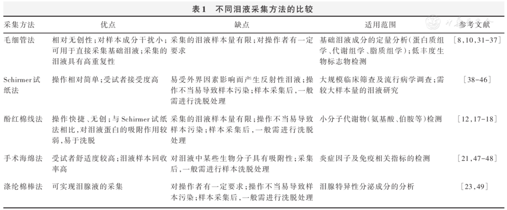
Table 1 Comparison of different tear collection methods
▼click to see the rest
References
[1] Vergouwen D, Schotting AJ, Endermann T, et al. Evaluation of pre-processing methods for tear fluid proteomics using proximity extension assays[J]. Sci Rep, 2023, 13(1)∶ 4433. DOI: 10.1038/s41598-023-31227-1.
▼click to see more references
[2] Zhou L, Beuerman RW. The power of tears: how tear proteomics research could revolutionize the clinic[J]. Expert Rev Proteomics, 2017, 14(3):189-191. DOI: 10.1080/14789450.2017.1285703.
[3] Kecskeméti G, Tóth-Molnár E, Janáky T, et al. An extensive study of phenol red thread as a novel non-invasive tear sampling technique for proteomics studies: comparison with two commonly used methods[J]. Int J Mol Sci, 2022, 23(15):8647. DOI: 10.3390/ijms23158647.
[4] Pieczyński J, Szulc U, Harazna J, et al. Tear fluid collection methods: review of current techniques[J]. Eur J Ophthalmol, 2021, 31(5):2245-2251. DOI: 10.1177/1120672121998922.
[5] Rentka A, Koroskenyi K, Harsfalvi J, et al. Evaluation of commonly used tear sampling methods and their relevance in subsequent biochemical analysis[J]. Ann Clin Biochem, 2017, 54(5):521-529. DOI: 10.1177/0004563217695843
[6] Wolffsohn JS, Benítez-Del-Castillo JM, Loya-Garcia D, et al. TFOS DEWS III: diagnostic methodology[J]. Am J Ophthalmol, 2025, 279:387-450. DOI: 10.1016/j.ajo.2025.05.033.
[7] Gijs M, van de Sande N, Bonnet C, et al. A comprehensive scoping review of methodological approaches and clinical applications of tear fluid biomarkers[J]. Prog Retin Eye Res, 2025, 106:101338. DOI: 10.1016/j.preteyeres.2025.101338.
[8] Blanco-Vázquez M, Vázquez A, Fernández I, et al. Inflammation-related molecules in tears of patients with chronic ocular pain and dry eye disease[J]. Exp Eye Res, 2022, 219:109057. DOI: 10.1016/j.exer.2022.109057.
[9] Frenia K, Fu Y, Beatty MA, et al. Precision in tear fluid biomarker discovery: quantitative proteomic profiling of small-volume, individual samples using capillary tube collection[J]. Biomedicines, 2025, 13(2):386. DOI: 10.3390/biomedicines13020386.
[10] Nokhoijav E, Guba A, Kumar A, et al. Metabolomic analysis of serum and tear samples from patients with obesity and type 2 diabetes mellitus[J]. Int J Mol Sci, 2022, 23(9):4534. DOI: 10.3390/ijms23094534.
[11] Harkness BM, Hegarty DM, Saugstad JA, et al. Experimental design considerations for studies of human tear proteins[J]. Ocul Surf, 2023, 28:58-78. DOI: 10.1016/j.jtos.2023.02.005.
[12] Saleh TA, McDermott B, Bates AK, et al. Phenol red thread test vs Schirmer’s test: a comparative study[J]. Eye (Lond), 2006, 20(8):913-915. DOI: 10.1038/sj.eye.6702052.
[13] Thormann K, Lüthi AS, Deniau F, et al. Dupilumab-associated ocular surface disease is characterized by a shift from Th2/Th17 toward Th1/Th17 inflammation[J]. Allergy, 2024, 79(4):937-948. DOI: 10.1111/all.16045.
[14] Tsai T, Alwees M, Rost A, et al. Changes of subjective symptoms and tear film biomarkers following Femto-LASIK[J]. Int J Mol Sci, 2022, 23(14):7512. DOI: 10.3390/ijms23147512.
[15] Gijs M, Adelaar TI, Vergouwen D, et al. Tear fluid inflammatory proteome analysis highlights similarities between keratoconus and allergic conjunctivitis[J]. Invest Ophthalmol Vis Sci, 2023, 64(15):9. DOI: 10.1167/iovs.64.15.9.
[16] Feret N, Ter Schiphorst S, Kindermans J, et al. Temporal analysis of tear fluid proteome reveals critical corneal repair events after photorefractive surgery[J]. Invest Ophthalmol Vis Sci, 2025, 66(13):49. DOI: 10.1167/iovs.66.13.49.
[17] Barmada A, Shippy SA. Quantifying sample collection and processing impacts on fiber-based tear fluid chemical analysis[J]. Transl Vis Sci Technol, 2020, 9(10):23. DOI: 10.1167/tvst.9.10.23.
[18] Avilov V, Zeng Q, Shippy SA. Threads for tear film collection and support in quantitative amino acid analysis[J]. Anal Bioanal Chem, 2016, 408(19):5309-5317. DOI: 10.1007/s00216-016-9624-7.
[19] Inic-Kanada A, Nussbaumer A, Montanaro J, et al. Comparison of ophthalmic sponges and extraction buffers for quantifying cytokine profiles in tears using Luminex technology[J]. Mol Vis, 2012, 18:2717-2725.
[20] Ma J, Sze YH, Bian JF, et al. Critical role of mass spectrometry proteomics in tear biomarker discovery for multifactorial ocular diseases (review)[J]. Int J Mol Med, 2021, 47(5):83. DOI: 10.3892/ijmm.2021.4916.
[21] Acera A, Rocha G, Vecino E, et al. Inflammatory markers in the tears of patients with ocular surface disease[J]. Ophthalmic Res, 2008, 40(6):315-321. DOI: 10.1159/000150445.
[22] Ji YW, Mittal SK, Hwang HS, et al. Lacrimal gland-derived IL-22 regulates IL-17-mediated ocular mucosal inflammation[J]. Mucosal Immunol, 2017, 10(5):1202-1210. DOI: 10.1038/mi.2016.119.
[23] Lee S, Kim E, Moon CE, et al. Amplified fluorogenic immunoassay for early diagnosis and monitoring of Alzheimer’s disease from tear fluid[J]. Nat Commun, 2023, 14(1):8153. DOI: 10.1038/s41467-023-43995-5.
[24] Fu R, Klinngam W, Heur M, et al. Tear proteases and protease inhibitors: potential biomarkers and disease drivers in ocular surface disease[J]. Eye Contact Lens, 2020, 46 Suppl 2:S70-S83. DOI: 10.1097/ICL.0000000000000641.
[25] Altman J, Jones G, Ahmed S, et al. Tear film microRNAs as potential biomarkers: a review[J]. Int J Mol Sci, 2023, 24(4):3694. DOI: 10.3390/ijms24043694.
[26] Nättinen J, Aapola U, Jylhä A, et al. Comparison of capillary and Schirmer strip tear fluid sampling methods using SWATH-MS proteomics approach[J]. Transl Vis Sci Technol, 2020, 9(3):16. DOI: 10.1167/tvst.9.3.16.
[27] Bruszel B, Tóth-Molnár E, Janáky T, et al. Sources of variance in human tear proteomic samples: statistical evaluation, quality control, normalization, and biological insight[J]. Int J Mol Sci, 2024, 25(3):1559. DOI: 10.3390/ijms25031559.
[28] Butovich IA. Tear film lipids[J]. Exp Eye Res, 2013, 117:4-27. DOI: 10.1016/j.exer.2013.05.010.
[29] Guyette N, Williams L, Tran MT, et al. Comparison of low-abundance biomarker levels in capillary-collected nonstimulated tears and washout tears of aqueous-deficient and normal patients[J]. Invest Ophthalmol Vis Sci, 2013, 54(5):3729-3737. DOI: 10.1167/iovs.12-11431.
[30] García-Porta N, Mann A, Sáez-Martínez V, et al. The potential influence of Schirmer strip variables on dry eye disease characterisation, and on tear collection and analysis[J]. Cont Lens Anterior Eye, 2018, 41(1):47-53. DOI: 10.1016/j.clae.2017.09.012.
[31] Kalsow CM, Reindel WT, Merchea MM, et al. Tear cytokine response to multipurpose solutions for contact lenses[J]. Clin Ophthalmol, 2013, 7:1291-1302. DOI: 10.2147/OPTH.S44642.
[32] Tummanapalli SS, Willcox M, Issar T, et al. Tear film substance P: a potential biomarker for diabetic peripheral neuropathy[J]. Ocul Surf, 2019, 17(4):690-698. DOI: 10.1016/j.jtos.2019.08.010.
[33] Eperon S, Berguiga M, Ballabeni P, et al. Total IgE and eotaxin (CCL11) contents in tears of patients suffering from seasonal allergic conjunctivitis[J]. Graefes Arch Clin Exp Ophthalmol, 2014, 252(9):1359-1367. DOI: 10.1007/s00417-014-2683-6.
[34] Ananthi S, Chitra T, Bini R, et al. Comparative analysis of the tear protein profile in mycotic keratitis patients[J]. Mol Vis, 2008, 14:500-507.
[35] Choy CK, Cho P, Chung WY, et al. Water-soluble antioxidants in human tears: effect of the collection method[J]. Invest Ophthalmol Vis Sci, 2001, 42(13):3130-3134.
[36] Srinivasan S, Joyce E, Boone A, et al. Tear lipocalin and lysozyme concentrations in postmenopausal women[J]. Ophthalmic Physiol Opt, 2010, 30(3):257-266. DOI: 10.1111/j.1475-1313.2010.00714.x.
[37] Catanese S, Khanna RK, Lefevre A, et al. Validation of metabolomic and lipidomic analyses of human tears using ultra-high-performance liquid chromatography tandem mass spectrometry[J]. Talanta, 2023, 253:123932. DOI: 10.1016/j.talanta.2022.123932.
[38] Wang Y, Xu Z, Gong Q, et al. The role of different tear volume detection methods in the evaluation and diagnosis of mild dry eye disease[J]. Transl Vis Sci Technol, 2022, 11(3):15. DOI: 10.1167/tvst.11.3.15.
[39] Matheis N, Grus FH, Breitenfeld M, et al. Proteomics differentiate between thyroid-associated orbitopathy and dry eye syndrome[J]. Invest Ophthalmol Vis Sci, 2015, 56(4):2649-2656. DOI: 10.1167/iovs.15-16699.
[40] Hu L, Zhang T, Ma H, et al. Discovering the secret of diseases by incorporated tear exosomes analysis via rapid-isolation system: iTEARS[J]. ACS Nano, 2022, 16(8):11720-11732. DOI: 10.1021/acsnano.2c02531.
[41] Tovar A, Gomez A, Serrano A, et al. Role of caspase-1 as a biomarker of ocular surface damage[J]. Am J Ophthalmol, 2022, 239:74-83. DOI: 10.1016/j.ajo.2022.01.020.
[42] Fatima A, Sanyal S, Jha GK, et al. The enigmatic world of tear extracellular vesicles (EVs)-exploring their role in ocular health and beyond[J]. FEBS Lett, 2025, 599(10):1346-1372. DOI: 10.1002/1873-3468.70004.
[43] Amorim M, Martins B, Caramelo F, et al. Putative biomarkers in tears for diabetic retinopathy diagnosis[J]. Front Med (Lausanne), 2022, 9:873483. DOI: 10.3389/fmed.2022.873483.
[44] Hamm-Alvarez SF, Janga SR, Edman MC, et al. Tear cathepsin S as a candidate biomarker for Sjögren’s syndrome[J]. Arthritis Rheumatol, 2014, 66(7):1872-1881. DOI: 10.1002/art.38633.
[45] Pieragostino D, Agnifili L, Fasanella V, et al. Shotgun proteomics reveals specific modulated protein patterns in tears of patients with primary open angle glaucoma naïve to therapy[J]. Mol Biosyst, 2013, 9(6):1108-1116. DOI: 10.1039/c3mb25463a.
[46] Park M, Jung H, Jeong Y, et al. Plasmonic Schirmer strip for human tear-based gouty arthritis diagnosis using surface-enhanced Raman scattering[J]. ACS Nano, 2017, 11(1):438-443. DOI: 10.1021/acsnano.6b06196.
[47] López-Cisternas J, Castillo-Díaz J, Traipe-Castro L, et al. Use of polyurethane minisponges to collect human tear fluid[J]. Cornea, 2006, 25(3):312-318. DOI: 10.1097/01.ico.0000183531.25201.0d.
[48] Martínez R, Acera A, Soria J, et al. Allergic mediators in tear from children with seasonal and perennial allergic conjunctivitis[J]. Arch Soc Esp Oftalmol, 2011, 86(6):187-192. DOI: 10.1016/j.oftal.2011.01.002.
[49] Jung JH, Ji YW, Hwang HS, et al. Proteomic analysis of human lacrimal and tear fluid in dry eye disease[J]. Sci Rep, 2017, 7(1):13363. DOI: 10.1038/s41598-017-13817-y.
[50] Roden RK, Zuniga N, Wright JC, et al. Human tear film protein sampling using soft contact lenses[J]. Clin Proteomics, 2024, 21(1):23. DOI: 10.1186/s12014-024-09475-8.
[51] Yao YN, Di D, Yuan ZC, et al. Schirmer paper noninvasive microsampling for direct mass spectrometry analysis of human tears[J]. Anal Chem, 2020, 92(9):6207-6212. DOI: 10.1021/acs.analchem.9b05078.
[52] Zhao W, Li J, Xue Z, et al. A separation-sensing platform performing accurate diagnosis of jaundice in complex biological tear fluids[J]. Angew Chem Int Ed Engl, 2022, 61(29):e202205628. DOI: 10.1002/anie.202205628.