Authors:Wang Xufan, Bai Wen, Yao Jin
Corresponding author: Yao Jin, Email: dryaojin@126.com
Published:2026-09-10
DOI:10.3760/cma.j.cn115989-20260407-00164
ABSTRACT
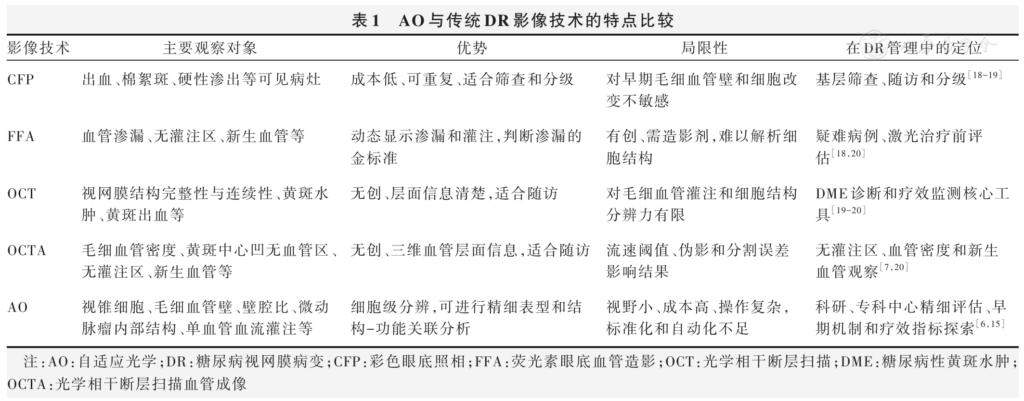
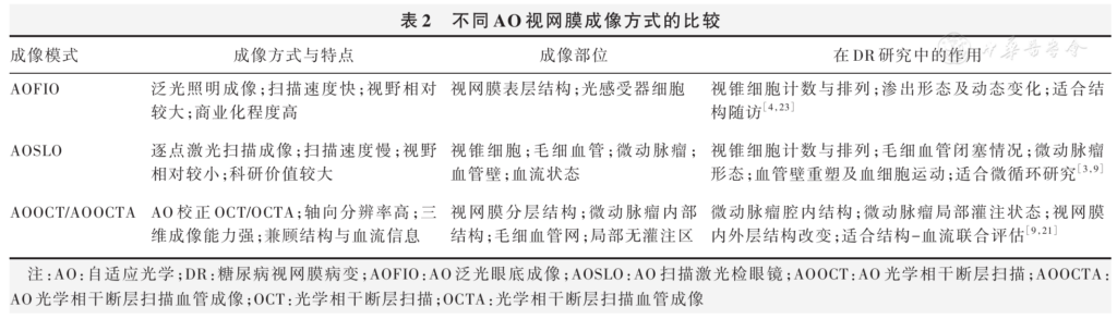
Diabetic retinopathy (DR) is a common microvascular complication in patients with diabetes. It is also an important cause of visual impairment in working-age adults. Color fundus photography, fundus fluorescein angiography, optical coherence tomography, and optical coherence tomography angiography are commonly used for DR screening, grading, and follow-up. These methods can show hemorrhage, leakage, edema, and nonperfusion areas. However, they are still limited in showing fine structures, including photoreceptors, capillary walls, and the internal structure of microaneurysms. Adaptive optics (AO) measures and corrects higher-order aberrations of the eye. It can improve the lateral resolution of retinal images and allow observation of early microvascular remodeling, local blood flow changes, and retinal neural damage at a near-cellular level. Recent studies suggest that AO is more suitable as a supplementary method after routine imaging rather than a primary screening tool. It can be used for detailed structural analysis, observation of structure-function relationships, and follow-up after treatment. This review summarizes the application of AO in DR, its complementary value with conventional imaging, and its current limitations.
KEYWORDS:
Diabetic retinopathy;Adaptive optics;Microvasculature;Photoreceptors;Retinal imaging;Multimodal imaging
COPYRIGHTS:
Copyright by Chinese Medical Association
No content published by the journals of Chinese Medical Association may be reproduced or abridged without authorization. Please do not use or copy the layout and design of the journals without permission.
All articles published represent the opinions of the authors, and do not reflect the official policy of the Chinese Medical Association or the Editorial Board, unless this is clearly specified.
Authors Info & Affiliations
Wang Xufan
The Affiliated Eye Hospital of Nanjing Medical University, Nanjing 210029, China
Bai Wen
The Affiliated Eye Hospital of Nanjing Medical University, Nanjing 210029, China
Yao Jin
The Affiliated Eye Hospital of Nanjing Medical University, Nanjing 210029, China
Figures & Tables
References click to collapse
[1] 孙书园, 徐莎,江珍红,等.糖尿病视网膜病变的发病机制与治疗研 究进展[J]. 中国药学杂志, 2026, 61(3)∶231-239. DOI: 10.11669/ cpj.2026.03.001. Sun SY, Xu S, Jiang ZH, et al. Recent progress of mechanisms and therapeutic approaches in diabetic retinopathy[J]. Chin Pharm J, 2026, 61(3)∶231-239. DOI: 10.11669/cpj.2026.03.001.
[2] 钱玖林,廖萱,兰长骏.自适应光学技术在眼科的应用进展[J].中华实 验眼科杂志, 2022, 40(3)∶284-288. DOI: 10.3760/cma.j.cn115989 20200801‐00550. Qian JL, Liao X, Lan CJ. Progress of application of adaptive optics in ophthalmology[J]. Chin J Exp Ophthalmol, 2022, 40(3)∶ 284-288. DOI: 10.3760/cma.j.cn115989‐20200801‐00550.
[3] 张剑平, 张美霞.自适应光学扫描技术在眼底检查中的应用进展[J]. 中华眼底病杂志, 2019, 35(6)∶ 621-623. DOI: 10.3760/cma. j.issn.1005‐1015.2019.06.025. Zhang JP, Zhang MX. The clinical applications of adaptive optics scanning laser ophthalmoscope[J]. Chin J Ocul Fundus Dis, 2019, 35(6)∶ 621-623. DOI: 10.3760/cma.j.issn.1005‐1015.2019.06.025.
[4] 张影, 张荟颖, 姚进.基于自适应光学的视网膜成像技术应用进展 [J]. 国 际 眼 科 杂 志, 2023, 23(12)∶ 1978-1982. DOI: 10.3980/j. issn.1672‐5123.2023.12.08. Zhang Y, Zhang HY, Yao J. Advancements in the application of adaptive optics‐based retinal imaging technology[J]. Int Eye Sci, 2023, 23(12)∶1978-1982. DOI: 10.3980/j.issn.1672‐5123.2023.12.08.
[5] 安广琪, 赵旭华,金学民,等.自适应光学成像在黄斑区光感受器细 胞观察中的研究进展[J]. 中华眼科杂志, 2025, 61(10)∶826-830. DOI: 10.3760/cma.j.cn112142‐20241106‐00498. An GQ, Zhao XH, Jin XM, et al. Advances in adaptive optical imaging of photoreceptor cell observation in the macular[J]. Chin J Ophthalmol, 2025, 61(10)∶ 826-830. DOI: 10.3760/cma. j. cn112142‐20241106 00498.
[6] Mirescu AE, Deleanu DG, Baltă G, et al. Adaptive optics imaging in diabetic retinopathy: a comprehensive review[J]. Rom J Ophthalmol, 2025, 69(3)∶299-309. DOI: 10.22336/rjo.2025.49.
[7] Vaughan M, Denmead P, Tay N, et al. How early can we detect diabetic retinopathy? A narrative review of imaging tools for structural assessment of the retina[J]. Graefes Arch Clin Exp Ophthalmol, 2025, 263(9)∶2413-2425. DOI: 10.1007/s00417‐025‐06828‐3.
[8] Grad J, Hatamnejad A, Pattathil N, et al. Photoreceptor characteristics in diabetic retinopathy vs controls using adaptive optics imaging: systematic review[J]. J Vitreoretin Dis, 2024∶ 24741264241286682. DOI: 10.1177/24741264241286682.
[9] Torm M, Pircher M, Bonnin S, et al. Detection of capillary abnormalities in early diabetic retinopathy using scanning laser ophthalmoscopy and optical coherence tomography combined with adaptive optics[J]. Sci Rep, 2024, 14(1)∶13450. DOI: 10.1038/s41598‐024‐63749‐7.
[10]Vaughan M, Tay N, Kalitzeos A, et al. Changes in waveguiding cone photoreceptors and color vision in patients with diabetes mellitus[J]. Invest Ophthalmol Vis Sci, 2024, 65(14)∶ 28. DOI: 10.1167/ iovs.65.14.28.
[11]Lai C, Su T, Cao J, et al. Retinal neurovascular impairment in full‐course diabetic retinopathy: the Guangdong diabetic retinopathy multiple‐omics study[J]. Invest Ophthalmol Vis Sci, 2024, 65(14)∶20. DOI: 10.1167/iovs.65.14.20.
[12]Matuszewski W, Szklarz M, Wołos‐Kłosowicz K, et al. High‐resolution imaging of cones and retinal arteries in patients with diabetes mellitus type 1 using adaptive optics (rtx1)[J]. Biomedicines, 2024, 12(4)∶863. DOI: 10.3390/biomedicines12040863.
[13]Parravano M, Fragiotta S, Costanzo E, et al. Metabolic, microvascular, and structural predictors of long‐term functional changes evaluated by multifocal electroretinogram in type 1 diabetes[J]. Biomedicines, 2024, 12(11)∶2614. DOI: 10.3390/biomedicines12112614.
[14]Pieczyński J, Berlińska A, Harazny JM. Retinal organisation and systemic vascular changes assessed by adaptive optics and doppler ultrasonography following anti‐VEGF therapy in patients with diabetic macular oedema[J]. Biomedicines, 2026, 14(1)∶ 124. DOI: 10.3390/ biomedicines14010124.
[15]Balas M, Issa M, Popovic MM, et al. Adaptive optics imaging in diabetic retinopathy: a prospective cohort study[J]. Retina, 2024, 44(7)∶ 1115-1123. DOI: 10.1097/IAE.0000000000004088.
[16]Balas M, Issa M, Popovic MM, et al. Correlation between photoreceptor and vascular parameters in diabetic retinopathy using adaptive optics [J]. Ophthalmic Surg Lasers Imaging Retina, 2025, 56(3)∶ 150-158. DOI: 10.3928/23258160‐20241015‐03.
[17]Su T, Lai C, Du Z, et al. Retinal photoreceptors and arterioles alterations detected using adaptive optics scanning laser ophthalmoscopy in diabetes and diabetic retinopathy[J]. Retina, 2026, 46(1)∶163-170. DOI: 10.1097/IAE.0000000000004647.
[18]Tran K, Pakzad‐Vaezi K. Multimodal imaging of diabetic retinopathy[J]. Curr Opin Ophthalmol, 2018, 29(6)∶ 566-575. DOI: 10.1097/ ICU.0000000000000524.
[19]Cole ED, Novais EA, Louzada RN, et al. Contemporary retinal imaging techniques in diabetic retinopathy: a review[J]. Clin Exp Ophthalmol, 2016, 44(4)∶289-299. DOI: 10.1111/ceo.12711.
[20]Nanegrungsunk O, Patikulsila D, Sadda SR. Ophthalmic imaging in diabetic retinopathy: a review[J]. Clin Exp Ophthalmol, 2022, 50(9)∶ 1082-1096. DOI: 10.1111/ceo.14170.
[21]Brunner E, Kunze L, Laidlaw V, et al. Improvements on speed, stability and field of view in adaptive optics OCT for anterior retinal imaging using a pyramid wavefront sensor[J]. Biomed Opt Express, 2024, 15(10)∶ 6098-6116. DOI: 10.1364/BOE.533451.
[22]Mujat M, Sampani K, Patel AH, et al. Cellular‐level analysis of retinal blood vessel walls based on phase gradient images[J]. Diagnostics (Basel), 2023, 13(22)∶3399. DOI: 10.3390/diagnostics13223399.
[23]Soliman MK, Sadiq MA, Agarwal A, et al. High‐resolution imaging of parafoveal cones in different stages of diabetic retinopathy using adaptive optics fundus camera[J/OL]. PLoS One, 2016, 11(4)∶ e0152788[2026-04-02]. https://pubmed. ncbi. nlm. nih. gov/27057752/. DOI: 10.1371/journal.pone.0152788.
[24]Sapoznik KA, Gast TJ, Carmichael‐Martins A, et al. Retinal arteriolar wall remodeling in diabetes captured with AOSLO[J]. Transl Vis Sci Technol, 2023, 12(11)∶16. DOI: 10.1167/tvst.12.11.16.
[25]AbdelAl O, Ashraf M, Sampani K, et al. “For Mass Eye and Ear Special Issue” adaptive optics in the evaluation of diabetic retinopathy[J]. Semin Ophthalmol, 2019, 34(4)∶ 189-197. DOI: 10.1080/08820538. 2019.1620794.
[26]Lombardo M, Parravano M, Serrao S, et al. Analysis of retinal capillaries in patients with type 1 diabetes and nonproliferative diabetic retinopathy using adaptive optics imaging[J]. Retina, 2013, 33(8)∶ 1630-1639. DOI: 10.1097/IAE.0b013e3182899326.
[27]Burns SA, Elsner AE, Chui TY, et al. In vivo adaptive optics microvascular imaging in diabetic patients without clinically severe diabetic retinopathy[J]. Biomed Opt Express, 2014, 5(3)∶ 961-974. DOI: 10.1364/BOE.5.000961.
[28]Sampani K, Mujat M, Patel AH, et al. Characterizing vascular wall and lumen caliber in eyes with diabetic retinopathy based on adaptive optics scanning laser ophthalmoscopy[J]. Diagnostics (Basel), 2024, 14(18)∶ 2020. DOI: 10.3390/diagnostics14182020.
[29]Baltă F, Cristescu IE, Mirescu AE, et al. Investigation of retinal microcirculation in diabetic patients using adaptive optics ophthalmoscopy and optical coherence angiography[J]. J Diabetes Res, 2022, 2022∶1516668. DOI: 10.1155/2022/1516668.
[30]Ueno Y, Iwase T, Goto K, et al. Association of changes of retinal vessels diameter with ocular blood flow in eyes with diabetic retinopathy[J]. Sci Rep, 2021, 11(1)∶4653. DOI: 10.1038/s41598‐021‐84067‐2.
[31]Huang BB, Fawzi AA. Hypertension likely drives arteriolar wall thickening in preclinical diabetic retinopathy while diabetes drives wall thickness in clinical retinopathy[J]. Transl Vis Sci Technol, 2024, 13(6)∶ 8. DOI: 10.1167/tvst.13.6.8.
[32]Luo T, Gast TJ, Vermeer TJ, et al. Retinal vascular branching in healthy and diabetic subjects[J]. Invest Ophthalmol Vis Sci, 2017, 58(5)∶ 2685-2694. DOI: 10.1167/iovs.17‐21653.
[33]Arichika S, Uji A, Murakami T, et al. Correlation of retinal arterial wall thickness with atherosclerosis predictors in type 2 diabetes without clinical retinopathy[J]. Br J Ophthalmol, 2017, 101(1)∶ 69-74. DOI: 10.1136/bjophthalmol‐2016‐309612.
[34]Zaleska‐Żmijewska A, Wawrzyniak ZM, Dąbrowska A, et al. Adaptive optics (rtx1) high‐resolution imaging of photoreceptors and retinal arteries in patients with diabetic retinopathy[J]. J Diabetes Res, 2019, 2019∶9548324. DOI: 10.1155/2019/9548324.
[35]Tam J, Dhamdhere KP, Tiruveedhula P, et al. Disruption of the retinal parafoveal capillary network in type 2 diabetes before the onset of diabetic retinopathy[J]. Invest Ophthalmol Vis Sci, 2011, 52(12)∶ 9257-9266. DOI: 10.1167/iovs.11‐8481.
[36]Chui TY, Pinhas A, Gan A, et al. Longitudinal imaging of microvascular remodelling in proliferative diabetic retinopathy using adaptive optics scanning light ophthalmoscopy[J]. Ophthalmic Physiol Opt, 2016, 36(3)∶290-302. DOI: 10.1111/opo.12273.
[37]Cristescu IE, Ochinciuc R, Balta F, et al. High‐resolution imaging of diabetic retinopathy lesions using an adaptive optics retinal camera[J]. Rom J Ophthalmol, 2019, 63(1)∶29-34.
[38]Huang BB, Fukuyama H, Burns SA, et al. Imaging the retinal vascular mural cells in vivo: elucidating the timeline of their loss in diabetic retinopathy[J]. Arterioscler Thromb Vasc Biol, 2024, 44(2)∶ 465-476. DOI: 10.1161/ATVBAHA.123.320169.
[39]Decker NL, Huang BB, Moonjely J, et al. Quantifying capillary pericytes in vivo links them to retinal vascular leakage and potentially albuminuria: a pilot study in diabetes[J]. Arterioscler Thromb Vasc Biol, 2025, 45(12)∶2277-2291. DOI: 10.1161/ATVBAHA.125.323278.
[40]Dubow M, Pinhas A, Shah N, et al. Classification of human retinal microaneurysms using adaptive optics scanning light ophthalmoscope fluorescein angiography[J]. Invest Ophthalmol Vis Sci, 2014, 55(3)∶ 1299-1309. DOI: 10.1167/iovs.13‐13122.
[41]Hafner J, Salas M, Scholda C, et al. Dynamic changes of retinal microaneurysms in diabetes imaged with in vivo adaptive optics optical coherence tomography[J]. Invest Ophthalmol Vis Sci, 2018, 59(15)∶ 5932-5940. DOI: 10.1167/iovs.18‐24573.
[42]Bernabeu MO, Lu Y, Abu‐Qamar O, et al. Estimation of diabetic retinal microaneurysm perfusion parameters based on computational fluid dynamics modeling of adaptive optics scanning laser ophthalmoscopy [J]. Front Physiol, 2018, 9∶989. DOI: 10.3389/fphys.2018.00989.
[43]Loganadane P, Delbosc B, Saleh M. Short‐term progression of diabetic hard exudates monitored with high‐resolution camera[J]. Ophthalmic Res, 2019, 61(1)∶3-9. DOI: 10.1159/000493858.
[44]Palochak C, Lee HE, Song J, et al. Retinal blood velocity and flow in early diabetes and diabetic retinopathy using adaptive optics scanning laser ophthalmoscopy[J]. J Clin Med, 2019, 8(8)∶1165. DOI: 10.3390/ jcm8081165.
[45]Czaja B, de Bouter J, Heisler M, et al. The effect of stiffened diabetic red blood cells on wall shear stress in a reconstructed 3D microaneurysm[J]. Comput Methods Biomech Biomed Engin, 2022, 25(15)∶1691-1709. DOI: 10.1080/10255842.2022.2034794.
[46]Raghavendra AJ, Damani A, Oechsli S, et al. Measurement of retinal blood flow precision in the human eye with multimodal adaptive optics imaging[J]. Biomed Opt Express, 2024, 15(8)∶ 4625-4641. DOI: 10.1364/BOE.524944.
[47]Lammer J, Prager SG, Cheney MC, et al. Cone photoreceptor irregularity on adaptive optics scanning laser ophthalmoscopy correlates with severity of diabetic retinopathy and macular edema[J]. Invest Ophthalmol Vis Sci, 2016, 57(15)∶ 6624-6632. DOI: 10.1167/ iovs.16‐19537.
[48]Lombardo M, Parravano M, Serrao S, et al. Investigation of adaptive optics imaging biomarkers for detecting pathological changes of the cone mosaic in patients with type 1 diabetes mellitus[J/OL]. PLoS One, 2016, 11(3)∶ e0151380[2026-04-02]. https://pubmed. ncbi. nlm. nih. gov/26963392/. DOI: 10.1371/journal.pone.0151380.
[49]Ro‐Mase T, Ishiko S, Omae T, et al. Association between alterations of the choriocapillaris microcirculation and visual function and cone photoreceptors in patients with diabetes[J]. Invest Ophthalmol Vis Sci, 2020, 61(6)∶1. DOI: 10.1167/iovs.61.6.1.
[50]Gu Q, Pan T, Cheng R, et al. Macular vascular and photoreceptor changes for diabetic macular edema at early stage[J]. Sci Rep, 2024, 14(1)∶20544. DOI: 10.1038/s41598‐024‐71286‐6.
[51]赵紫昱, 张钊君, 王绪泉,等.糖尿病视网膜病变光学成像与人工智 能评估技术的研究进展[J].科学通报,2026,71(19)∶4537-4557. Zhao ZY, Zhang ZJ, Wang XQ, et al. Research progress on optical imaging and artificial intelligence assessment technologies for diabetic retinopathy[J]. Chin Sci Bull, 2026, 71(19)∶4537-4557.
[52]Attiku Y, He Y, Nittala MG, et al. Current status and future possibilities of retinal imaging in diabetic retinopathy care applicable to low‐ and medium‐income countries[J]. Indian J Ophthalmol, 2021, 69(11)∶ 2968-2976. DOI: 10.4103/ijo.IJO_1212_21.
[53]宁岸, 叶夏笛, 杨建文,等.自适应光学眼底高分辨成像技术与应用 [J]. 光 电 工 程 , 2025, 52(12)∶ 139-162. DOI: 10.12086/ oee.2025.250360. Ning A, Ye XD, Yang JW, et al. Techniques and applications of adaptive optics‐based high‐resolution fundus imaging[J]. Opto‐Electron Eng, 2025, 52(12)∶139-162. DOI: 10.12086/oee.2025.250360.