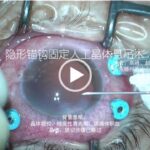
Minimally invasive anchoring hook fixation for intraocular lens suspension Surgeons: Gao Feng, Zhao Ruiling Tengzhou Central People’s Hospital, Zaozhuang 277500, Shandong Province, China Main steps: Use a 30G needle to thread the polypropylene suture through the 2.2 mm clear corneal incision. Fix the two intraocular lens loops sequentially and adjust the intraocular lens´s position before […]
Category: Media Center
Single-suture single-knot ab externo cyclopexy for post-traumatic cyclodialysis cleft
Surgeon:Xie Zhenggao Department of Ophthalmology, Nanjing Drum Tower Hospital, the Affiliated Hospital of Nanjing University Medical School Main steps: A laminar scleral flap incision parallel to the limbus was made 3.0 mm behind the limbus, ranging from 11:30 to 3:30. A 10-0 polypropylene straight needle docked into a 26-gauge needle was inserted into the anterior […]
Phacoemulsification under the intraocular lens protection
Phacoemulsification under the intraocular lens protection Main steps: Enlarge the main incision to 3.2 mm after water separation and water stratification. Implant the foldable intraocular lens (IOL) into the anterior chamber. Left one loop of the IOL in the main incision to stabilize the IOL. Perform phacoemulsification to extract the lens nucleus and the cortex […]
Human amniotic membrane plugging
Human amniotic membrane plugging Surgeon: Wang Zhaoyang Department of Ophthalmology, Shanghai Ninth People’s Hospital, Shanghai Jiao Tong University School of Medicine, Shanghai 200011, China Wang Zhaoyang now works at the Department of Ophthalmology, Shanghai Tenth People’s Hospital Affiliated to Tongji University, Shanghai 200072, China Main steps: The human amniotic membrane (hAM) patch was […]
Phacoemulsification combined with gonioscopy-assited angle plasty
Phacoemulsification combined with gonioscopy-assited angle plasty Intraoperatively, a clear corneal incision was made in the superior or superior temporal region. The viscoelastic was injected into the anterior chamber. Lens capsule rupture was performed. Liquid-liquid fractionation was performed with balanced salt solution. The lens nucleus was ultrasonically emulsified, and the lens cortex was aspirated with an […]
Phacoemulsification combined with intraocular lens implantation plus goniosynechialysis and goniotomy for advanced primary angle-closure glaucoma
Phacoemulsification combined with intraocular lens implantation plus goniosynechialysis and goniotomy for advanced primary angle-closure glaucoma The surgery was performed by Prof. Chen Weirong and Prof. Zhang Xiulan from Zhongshan Ophthalmic Center of Sun Yat-sen University. The right eye of patient received the operation. The phacoemulsification combined with intraocular lens implantation was finished by Prof. Chen. […]
Vitrectomy combined with internal limiting membrane peeling
Vitrectomy combined with internal limiting membrane peeling Application of standard three-channel 23G; posterior vitreous removal; peripheral vitreous removal with a scleral depressor; vitreous excision around the hole; indocyanine green staining; internal limiting membrane peeling around the macular hole; closure of degenerative area by laser; gas-liquid exchange; macular hole closure by laser; silicone oil tamponade.
Intraocular corrective lens implantation
Intraocular corrective lens implantation Placed in the ejector, the intraocular corrective lens (ICL) was pushed into the anterior chamber from the main incision and unfolded naturally. Then the four loops of ICL were adjusted to the posterior surface of the iris and fixed at the ciliary sulcus, and the residual viscoelastics was rinsed out of […]
Ultrasound cycloplasty for uncontrolled intraocular pressure after glaucoma surgery
Ultrasound cycloplasty for uncontrolled intraocular pressure after glaucoma surgery After draping patient for surgery, the retrobulbar injection of 3.5 ml of 2% lidocaine hydrochloride injection was performed to achieve anesthesia. Probe inserted in the coupling cone was fixated on the patient’s eye, then the negative pressure was started and the system detection was finished. The […]
Non-penetrating trabecular surgery combined with suture trabeculectomy for primary congenital glaucoma
Non-penetrating trabecular surgery combined with suture trabeculectomy for primary congenital glaucoma The patient was a 2-month-old boy with primary congenital glaucoma. The preoperative intraocular pressure was 28 mmHg, and corneal diameter was 13 mm, cup-to-disc ratio was 0.7. The canal of Schlemm was opened via non-penetrating trabecular surgery, then the cannulation with the twisted 6-0 […]
