Authors:Gu Liang, Gao Fengjie, Guan Huaijin, Ji Min
Corresponding author:Ji Min, Email:amyji1234@hotmail.com
Published:2026-08-10
DOI:10.3760/cma.j.cn115989-20250303-00063
ABSTRACT
Objective To analyze the clinical and genetic characteristics of a family with Waardenburg syndrome type Ⅰ (WS1) .
Methods A pedigree investigation was conducted. Clinical data from 3 generations of 7 people from a family with WS1 in the Nantong area of China, Jiangsu Province, who visited the Department of Ophthalmology at the Affiliated Hospital of Nantong University in April 2024, including 3 patients. Proband and her parents underwent comprehensive ophthalmic examinations, including slit-lamp examination, color fundus photography, optical coherence tomography, anterior segment photography, pure-tone audiometry, and general physical examination. Peripheral blood samples were obtained for whole-exome sequencing and subsequent data analysis. The pathogenic variants from members were analyzed according to the ACMG guidelines. This study adhered to the Declaration of Helsinki and was approved by the institutional Ethical Committee Review Board of Affiliated Hospital of Nantong University (No. 2024-L080). All subjects were informed of the purpose of this study and signed informed consent forms.
Results The proband, a 12-year-old girl, presented with dystopia canthorum, wide nasion, thick and connected eyebrows, bright blue irises, and diffuse hypopigmentation of the fundus with exposure of choroidal vessels in both eyes. The choroid was significantly thickened in both eyes, reaching a thickness of 509 µm in the right eye and 514 µm in the left eye. She also had moderate hearing loss in the left ear and severe hearing loss in the right ear. Physical examination of the proband’s mother showed premature gray hair, lateral deviation of the inner canthus of both eyes, wide nasion, and thick eyebrows. The left eye iris was bright blue, and the fundus was diffusely hypomelanotic with choroidal vessels exposed, showing a sunset glow-like fundus. The left eye choroid was significantly thickened to 558 µm. No obvious abnormalities were noted in the right eye. Audiometric testing revealed severe impairment in both ears. Whole exome sequencing showed that the proband and her mother carried a heterozygous nonsense variation c.667C>T (p.Arg223Ter) in the PAX3 gene, which caused premature termination of protein translation. This variant is classified as pathogenic according to ACMG guidelines. The inheritance pattern in this family is consistent with autosomal dominant inheritance.
Conclusions The c.667C>T mutation in the PAX3 gene is a pathogenic variant site in this WS1 family. The special manifestation of iris heterochromia accompanied by ipsilateral choroid thickening in this family has been reported for the first time in WS1.
KEYWORDS:
Waardenburg syndrome type Ⅰ;PAX3 gene ;Heterochromic iris;Choroidal thickening;c.667C>T (p.Arg223Ter)
COPYRIGHTS:
Copyright by Chinese Medical Association
No content published by the journals of Chinese Medical Association may be reproduced or abridged without authorization. Please do not use or copy the layout and design of the journals without permission.
All articles published represent the opinions of the authors, and do not reflect the official policy of the Chinese Medical Association or the Editorial Board, unless this is clearly specified.
Authors Info & Affiliations
Gu Liang
Department of Ophthalmology, Affiliated Hospital of Nantong University, Nantong 226001, China
Gao Fengjie
Department of Ophthalmology, Affiliated Hospital of Nantong University, Nantong 226001, China
Guan Huaijin
Department of Ophthalmology, Affiliated Hospital of Nantong University, Nantong 226001, China
Ji Min
Department of Ophthalmology, Affiliated Hospital of Nantong University, Nantong 226001, China
Figures & Tables
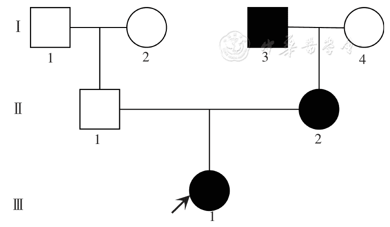
Figure 1 Pedigree map of type Ⅰ Waardenburg syndrome □: normal male; ○: normal female; ■: affected male; ●: affected female; ↗: proband
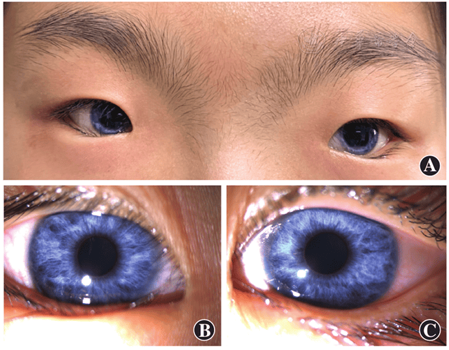
Figure 2 Facial and ocular clinical features of the proband The proband exhibited lateral deviation of the inner canthi of both eyes, a wide nasion, thick and prominent eyebrows, and vivid blue irises A: Face B: Right eye C: Left eye
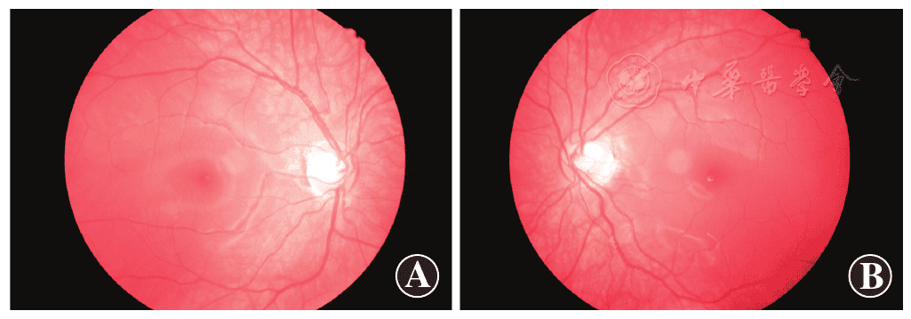
Figure 3 Fundus photographs of the proband’s eyes The retinas of both eyes appeared flat, exhibiting diffuse hypopigmentation and exposed choroidal vessels, which resulted in a sunset-like glow of the fundus A: Right eye B: Left eye
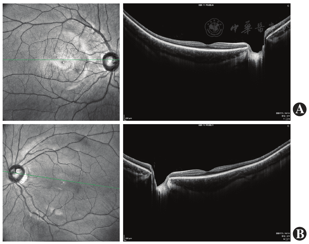
Figure 4 Optical coherence tomography images of the macular region in both eyes of the proband Significant thickening of the choroidal layer in both eyes A: Right eye B: Left eye
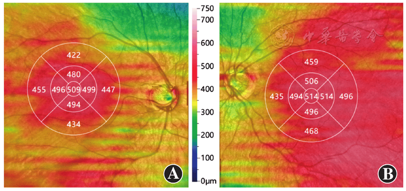
Figure 5 Comparison of choroidal thickness measured by optical coherence tomography angiography between the two eyes of the proband The choroidal thickness was 509 μm in the right eye and 514 μm in the left eye of the proband A: Right eye B: Left eye
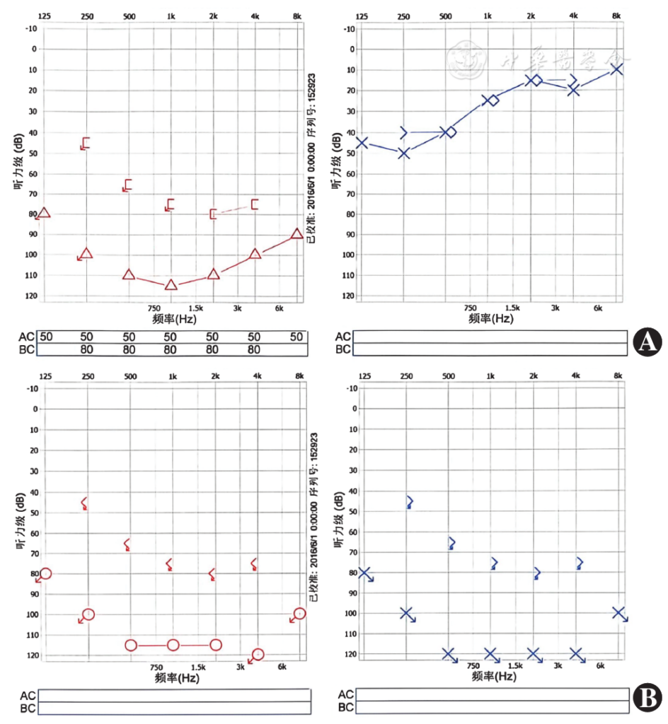
Figure 6 Binaural pure tone audiometry results of the proband and her mother A: The proband showed severe impairment in the right ear and moderate impairment in the left ear B: The proband’s mother showed bilateral severe impairment
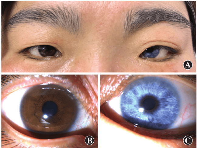
Figure 7 Facial and ocular clinical features of the proband’s mother Notable characteristics included bilateral lateral canthus, wide nasion, thick eyebrows, and a bright blue iris in the left eye of the proband’s mother A: Face B: Right eye C: Left eye
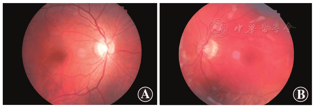
Figure 8 Fundus photography of the proband’s mother The fundus of both eyes of the proband’s mother appeared flat, exhibiting diffuse hypopigmentation and exposed choroidal vessels, which resulted in a sunset-like glow of the fundus A: Right eye B: Left eye
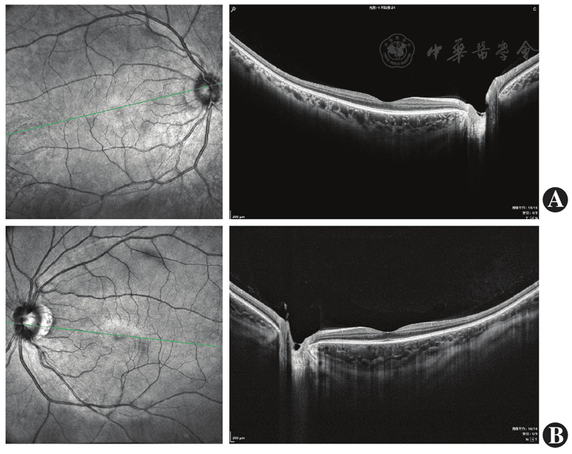
Figure 9 Optical coherence tomography examination of the binocular macula of the proband’s mother Significant thickening of the choroidal layer in the left eye A: Right eye B: Left eye
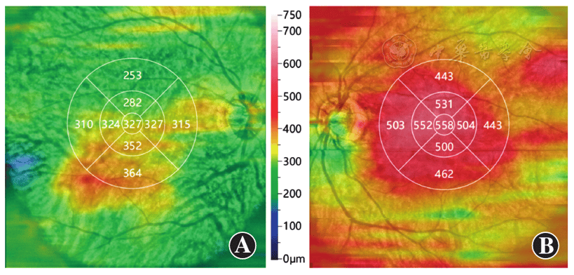
Figure 10 Comparison of choroidal thickness between the two eyes of the proband’s mother under optical coherence tomography angiography The left eye of proband’s mother exhibited a choroidal thickness of 558 μm, while the right eye had a choroidal thickness of 327 μm A: Right eye B: Left eye
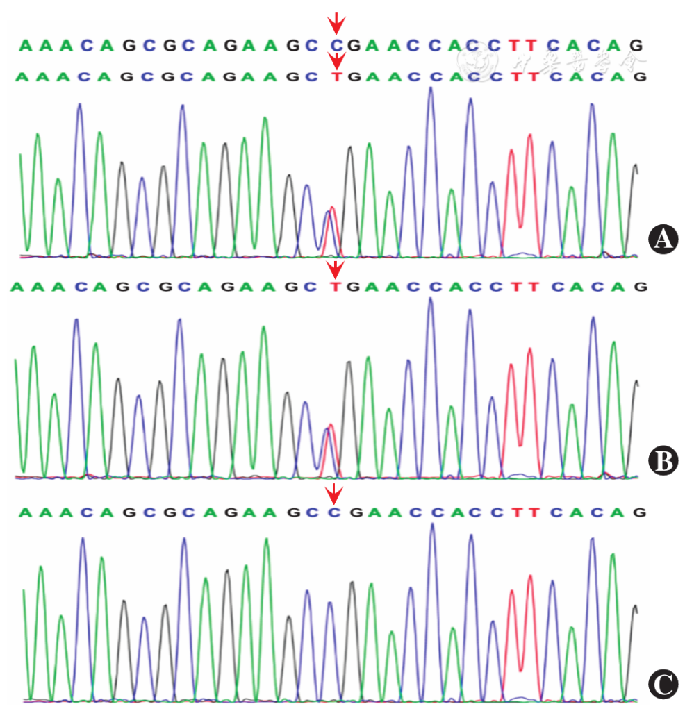
Figure 11 Sanger sequencing results A: Heterozygous mutation (c.667C>T) in the proband B: Heterozygous mutation (c.667C>T) in the proband’s mother C: Wild-type sequence in the proband’s father The arrow pointed to the variant site
References click to collapse
[1] Ramakrishnan IL, Taksande A. Waardenburg syndrome [J]. Pan Afr Med J, 2023, 45:23. DOI: 10.11604/pamj.2023.45.23.38829.
[2] Agrawal R, Walia S. Waardenburg syndrome type 1 [J]. Indian J Ophthalmol, 2022, 70 (7):2679‑2681. DOI: 10.4103/ijo.IJO_3003_21.
[3] Pingault V, Ente D, Dastot‑Le Moal F, et al. Review and update of mutations causing Waardenburg syndrome [J]. Hum Mutat, 2010, 31 (4):391‑406. DOI: 10.1002/humu.21211.
[4] Wang J, Li S, Xiao X, et al. PAX3 mutations and clinical characteristics in Chinese patients with Waardenburg syndrome type 1 [J]. Mol Vis, 2010, 16:1146‑1153.
[5] Farrer LA, Grundfast KM, Amos J, et al. Waardenburg syndrome (WS) type Ⅰ is caused by defects at multiple loci, one of which is near ALPP on chromosome 2: first report of the WS consortium [J]. Am J Hum Genet, 1992, 50 (5):902‑913.
[6] Kingdom R, Wright CF. Incomplete penetrance and variable expressivity: from clinical studies to population cohorts [J]. Front Genet, 2022, 13:920390. DOI: 10.3389/fgene.2022.920390.
[7] 张彩虹,温馨,王喜悦,等. PAX3 基因突变的 Waardenburg 综合征家系分析 [J]. 中国耳鼻咽喉颅底外科杂志,2024, 30 (4):56‑62. DOI: 10.11798/j.issn.1007‑1520.202423327.
Zhang CH, Wen X, Wang XY, et al. PAX3 gene mutation in a family with Waardenburg syndrome [J]. Chin J Otorhinolaryngol Skull Base Surg, 2024, 30 (4):56‑62. DOI: 10.11798/j.issn.1007‑1520.202423327.
[8] 曲春燕,赵敏,李俊,等。一个 Waardenburg 综合征患儿家系的 PAX3 基因突变分析 [J]. 中华耳科学杂志,2015, 13 (3):480‑483. DOI: 10.3969/j.issn.1672‑2922.2015.03.021.
Qu CY, Zhao M, Li J, et al. Genetic analysis of PAX3 gene in a Waardenburg syndrome family [J]. Chin J Otol, 2015, 13 (3):480‑483. DOI: 10.3969/j.issn.1672‑2922.2015.03.021.
[9] Bocangel M, Melo US, Alves LU, et al. Waardenburg syndrome: novel mutations in a large Brazilian sample [J]. Eur J Med Genet, 2018, 61 (6):348‑354. DOI: 10.1016/j.ejmg.2018.01.012.
[10] 张华,冯永. PAX3 基因对神经嵴发育的调控及其在 Waardenburg 综合征发病中的作用研究 [J]. 听力学及言语疾病杂志,2014, 22 (2):212‑215. DOI: 10.3969/j.issn.1006‑7299.2014.02.028.
[11] Udagawa T, Takahashi E, Tatsumi N, et al. Loss of Pax3 causes reduction of melanocytes in the developing mouse cochlea [J]. Sci Rep, 2024, 14 (1):2210. DOI: 10.1038/s41598‑024‑52629‑9.
[12] Chen K, Zhan Y, Wu X, et al. Germinal mosaicism of PAX3 mutation caused Waardenburg syndrome type I [J]. Int J Pediatr Otorhinolaryngol, 2018, 104:200‑204. DOI: 10.1016/j.ijporl.2017.11.011.
[13] Matsunaga T, Mutai H, Namba K, et al. Genetic analysis of PAX3 for diagnosis of Waardenburg syndrome type Ⅰ[J]. Acta Otolaryngol, 2013, 133 (4):345‑351. DOI: 10.3109/00016489.2012.744470.
[14] Baldwin CT, Lipsky NR, Hoth CF, et al. Mutations in PAX3 associated with Waardenburg syndrome type Ⅰ[J]. Hum Mutat, 1994, 3 (3):205‑211. DOI: 10.1002/humu.1380030306.
[15] Jalilian N, Tabatabaiefar MA, Farhadi M, et al. Molecular and clinical characterization of Waardenburg syndrome type Ⅰ in an Iranian cohort with two novel PAX3 mutations [J]. Gene, 2015, 574 (2):302‑307. DOI: 10.1016/j.gene.2015.08.023.
[16] Pandya A, Xia XJ, Landa BL, et al. Phenotypic variation in Waardenburg syndrome: mutational heterogeneity, modifier genes or polygenic background?[J]. Hum Mol Genet, 1996, 5 (4):497‑502. DOI: 10.1093/hmg/5.4.497.
[17] Yang SZ, Cao JY, Zhang RN, et al. Nonsense mutations in the PAX3 gene cause Waardenburg syndrome type Ⅰ in two Chinese patients [J]. Chin Med J (Engl), 2007, 120 (1):46‑49.
[18] Rishi P, Multani P, Prasan VV, et al. Choroidal thickness in Waardenburg syndrome [J]. GMS Ophthalmol Cases, 2019, 9:Doc22. DOI: 10.3205/oc000111.
[19] Choudhry N, Rao RC. Multimodal ultrawide‑field imaging features in Waardenburg syndrome [J]. Ophthalmic Surg Lasers Imaging Retina, 2015, 46 (6):670‑673. DOI: 10.3928/23258160‑20150610‑12.
[20] 蒋卓远,查艳,石小峰,等。神经嵴细胞和神经嵴病及其致病机制的研究进展 [J]. 遗传,2022, 44 (2):117‑134. DOI: 10.16288/j.yczz.21‑253.
Jiang ZY, Zha Y, Shi XF, et al. Research progress on neural crest cells and neurocristopathies and its pathogenesis [J]. Hereditas (Beijing), 2022, 44 (2):117‑134. DOI: 10.16288/j.yczz.21‑253.
[21] 李晶,马忠旭。神经嵴发育异常相关眼病 [J]. 国际眼科杂志,2024, 24 (1):53‑57. DOI: 10.3980/j.issn.1672‑5123.2024.1.10.
Li J, Ma ZX. Eye diseases associated with developmental abnormality of neural crest [J]. Int Eye Sci, 2024, 24 (1):53‑57. DOI: 10.3980/j.issn.1672‑5123.2024.1.10.
[22] He SJ, Stevens G, Braithwaite AW, et al. Transfection of melanoma cells with antisense PAX3 oligonucleotides additively complements cisplatin‑induced cytotoxicity [J]. Mol Cancer Ther, 2005, 4 (6):996‑1003. DOI: 10.1158/1535‑7163.MCT‑04‑0252.
[23] Ulrich E, Kistenmacher S, Martin G, et al. PAX3 expression patterns in ocular surface melanocytes [J]. Sci Rep, 2025, 15 (1):12472. DOI: 10.1038/s41598‑025‑90318‑3.
[24] 张华,常尚揆,冯永,等. PAX3 对靶基因 MITF 转录活性调控的实验研究 [J]. 听力学及言语疾病杂志,2015, 23 (5):505‑509. DOI: 10.3969/j.issn.1006‑7299.2015.05.015.
Zhang H, Chang SK, Feng Y, et al. Experimental studies on regulation of PAX3 on transcriptional activities of target gene MITF [J]. J Audiol Speech Pathol, 2015, 23 (5):505‑509. DOI: 10.3969/j.issn.1006‑7299.2015.05.015.
[25] Liang X, Dong Z, Bin W, et al. PAX3 promotes proliferation of human glioma cells by WNT/β‑catenin signaling pathways [J]. J Mol Neurosci, 2019, 68 (1):66‑77. DOI: 10.1007/s12031‑019‑01283‑2.
[26] 李云晓,杨慈清,林俊堂. Pax3 和 Pax7 在胚胎发育早期及神经嵴形成中的作用 [J]. 生物学杂志,2021, 38 (2):100‑104. DOI: 10.3969/j.issn.2095‑1736.2021.02.100.
Li YX, Yang CQ, Lin JT. The role of Pax3 and Pax7 in early embryonic development and neural crest formation [J]. J Biol, 2021, 38 (2):100‑104. DOI: 10.3969/j.issn.2095‑1736.2021.02.100.