Authors:Expert Workgroup of Expert consensus on the specification and operation of optical coherence tomography/optical coherence tomography angiography examination in experimental animals (2026), Ophthalmic Imaging and Intelligent Medicine Branch of Chinese Medicine Education Association, Ophthalmology Committee of International Association of Translational Medicine, Ophthalmology Committee of International Association of Intelligent Medicine, Chinese Ophthalmic Imaging Study Group
Corresponding authors: Shao Yi, Email: freebee99@163.com; Ji Dan, Email:jidan222@163.com; Yan Biao,Email: yanbiao1982@126.com
Published:2026-08-10
DOI: 10.3760/cma.j.cn115989-20241108-00306
ABSTRACT
Optical coherence tomography (OCT) and optical coherence tomography angiography (OCTA) have rapidly advanced over the past two decades as non-contact imaging techniques for tomographic analysis of intraocular tissue structures. Their applications in diagnosing and studying retinal diseases and glaucoma have revealed their unique clinical value. With the continuous innovation of technology, there are many brands of clinical instruments based on OCT/OCTA, and the iterative update is fast, so we need to improve the application level. In the field of experimental animal research, challenges arise due to physiological differences among various species of experimental animals, anatomical variations between the animals and the human eye, and limitations imposed by distinct research objectives. Moreover, anterior segment and retinal OCT/OCTA technologies represent high-resolution three-dimensional optical detection methods in vivo, posing difficulties for existing clinical instruments to meet the rigorous scientific and accuracy standards demanded by scientific research and efficacy evaluations. At the same time, the complexity of animal experiments, the diversity of scientific research tasks, and the constraints such as limited training time, insufficient experience in instrument operation, compared to clinical settings can lead to suboptimal OCT/OCTA outcomes for some research. To address this practical challenge, this consensus summarizes the technical principles, operation procedures and experimental precautions related to OCT/OCTA for the anterior segment and retina in common experimental animals. It also outlines the reference standards for corneal, retinal, and microvascular indicators across different animal models using OCT/OCTA technology, along with the practical applications of qualitative and quantitative analyses in ophthalmic corneal and retinal research.
KEYWORDS:
Optical coherence tomography;Optical coherence tomography angiography;Experimental animals;Operating specifications;Expert consensus
COPYRIGHTS:
Copyright by Chinese Medical Association
No content published by the journals of Chinese Medical Association may be reproduced or abridged without authorization. Please do not use or copy the layout and design of the journals without permission.
All articles published represent the opinions of the authors, and do not reflect the official policy of the Chinese Medical Association or the Editorial Board, unless this is clearly specified.
Authors Info & Affiliations
Expert Workgroup of Expert consensus on the specification and operation of optical coherence tomography/optical coherence tomography angiography examination in experimental animals (2026)
Ophthalmic Imaging and Intelligent Medicine Branch of Chinese Medicine Education Association
Ophthalmology Committee of International Association of Translational Medicine
Ophthalmology Committee of International Association of Intelligent Medicine
Chinese Ophthalmic Imaging Study Group
Shao Yi
Department of Ophthalmology, Shanghai General Hospital, Shanghai Jiao Tong University School of Medicine, National Clinical Research Center for Eye Diseases, Shanghai 200080, China
Ji Dan
Sichuan Academy of Medical Sciences, Sichuan Provincial People’s Hospital, Chengdu 610072, China
Yan Biao
Department of Ophthalmology, Shanghai General Hospital, Shanghai Jiao Tong University School of Medicine, National Clinical Research Center for Eye Diseases, Shanghai 200080, China
Figures & Tables
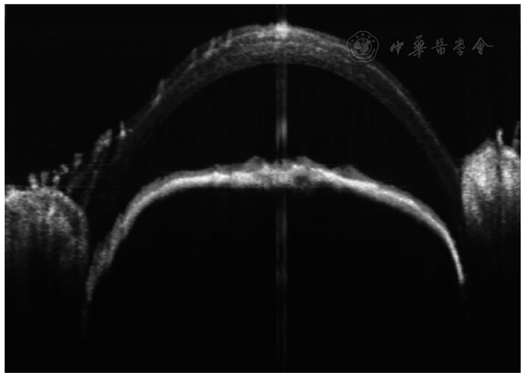
Figure 1 Non-standard corneal OCT images of C57BL/6 mice OCT: optical coherence tomography
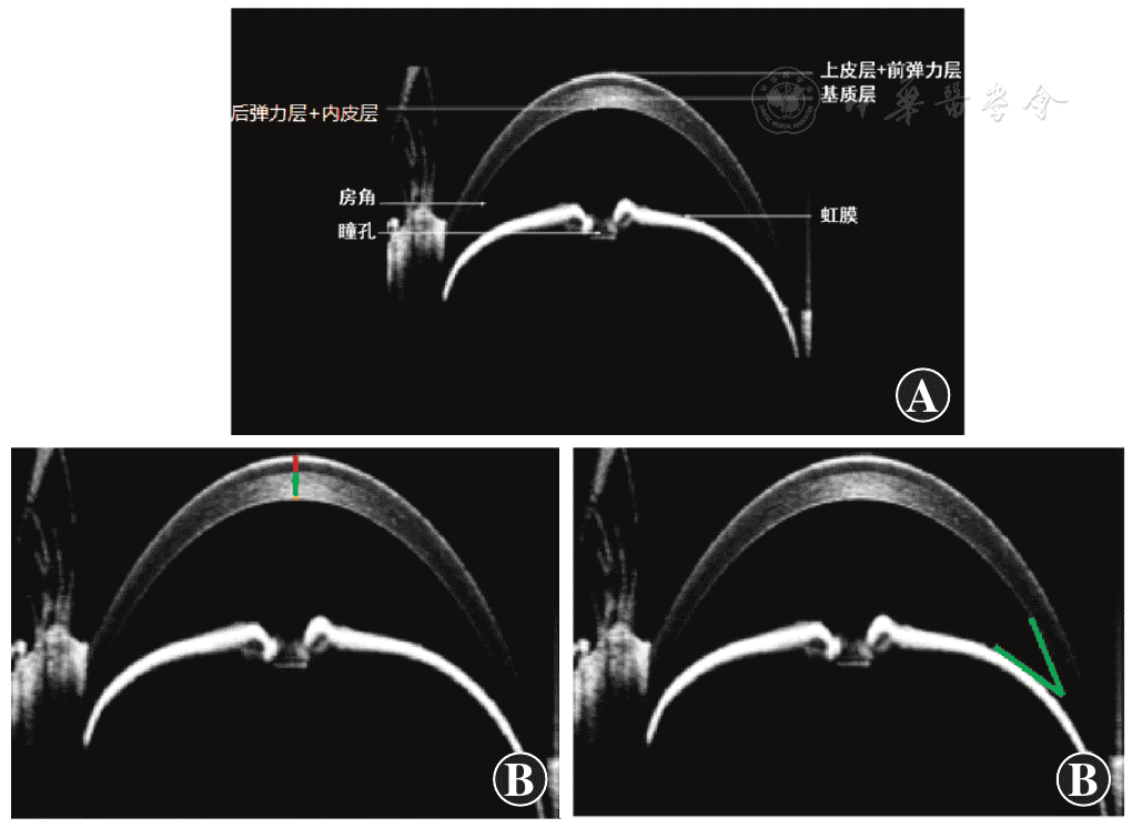
Figure 2 Corneal OCT image of normal C57BL/6 mice A: Corneal OCT image hierarchical annotation B: Corneal OCT thickness point-to-point hierarchical measurement diagram C: Corneal OCT chamber angle measurement diagram (chamber angle angle 30°) OCT: optical coherence tomography
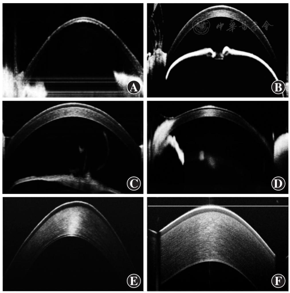
Figure 3 Corneal OCT images of normal adult animals in different species A: Zebrafish B: C57BL/6 mouse C: SD rat D: Guinea pig E: New Zealand white rabbit F: Pig OCT: optical coherence tomography
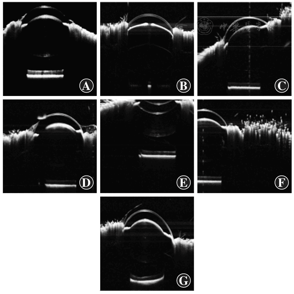
Figure 4 OCT measurement images of eye axis length in normal C57BL/6 mice A: High-quality image of eye axis measurement by OCT B: Low-quality image caused by not performing pupil dilation or incomplete pupil dilation C: Low-quality image caused by eyeball orientation not facing the lens D: Low-quality image caused by occlusion E: Low-quality image caused by failure to reach the optimal focus position F: Low-quality image caused by eyeball not being in the scan area G: Low-quality image caused by ocular surface fluid not being cleaned OCT: optical coherence tomography

Figure 5 Normal adult C57BL/6 mouse retinal OCT images A: Retinal OCT layer map B: Retinal OCT thickness measurement model map C: Low-quality retinal OCT image OCT: optical coherence tomography
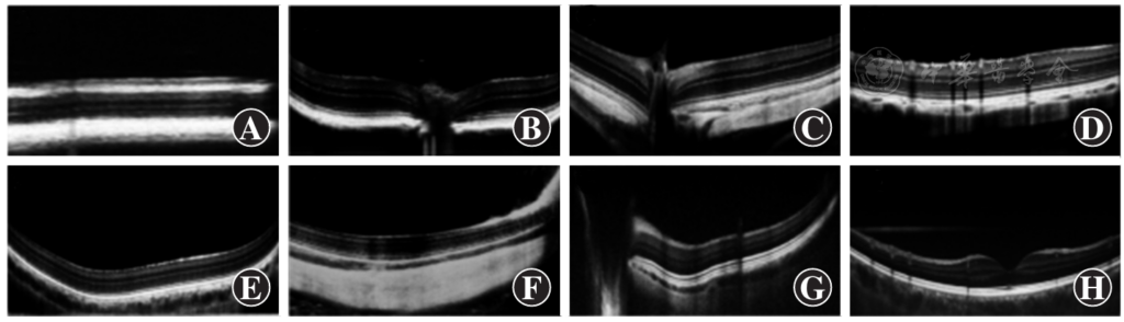
Figure 6 Retinal OCT images of experimental animals in different species A: Zebrafish B: C57BL/6 mouse C: SD rat D: BN rat E: Guinea pig F: New Zealand white rabbit G: pig H: rhesus monkey OCT: optical coherence tomography
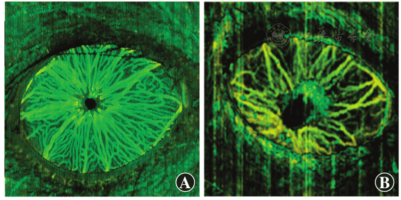
Figure 7 OCTA images of normal C57BL/6 mouse iris A: High-quality OCTA of iris B: Low-quality OCTA image of iris with motion artifacts and signal noise OCTA: optical coherence tomography angiography
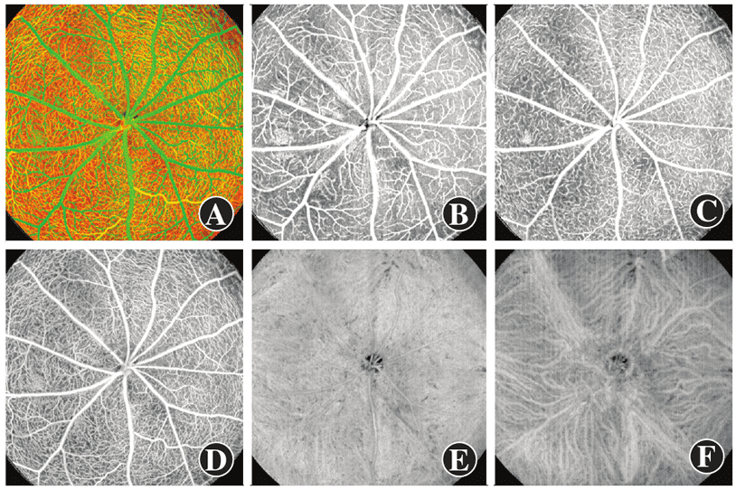
Figure 8 OCTA images of normal adult C57BL/6 mouse retina and choroid A: Retinal color map B: Gray map of nerve fiber layer C: Gray map of inner plexiform layer D: Gray map of outer plexiform layer E: Gray map of superficial choroid F: Gray map of deep choroid OCTA: optical coherence tomography angiography
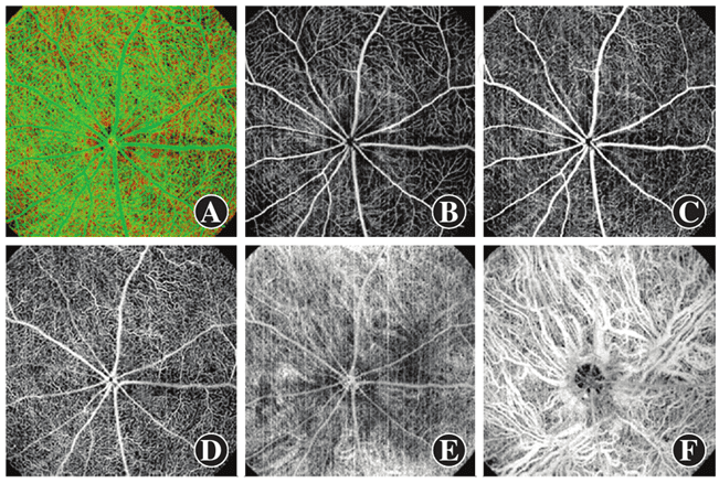
Figure 9 OCTA images of SD rat retina and choroid A: Retinal color map B: Gray map of nerve fiber layer C: Gray map of inner plexiform layer D: Gray map of outer plexiform layer E: Gray map of superficial choroid F: Gray map of deep choroid OCTA: optical coherence tomography angiography
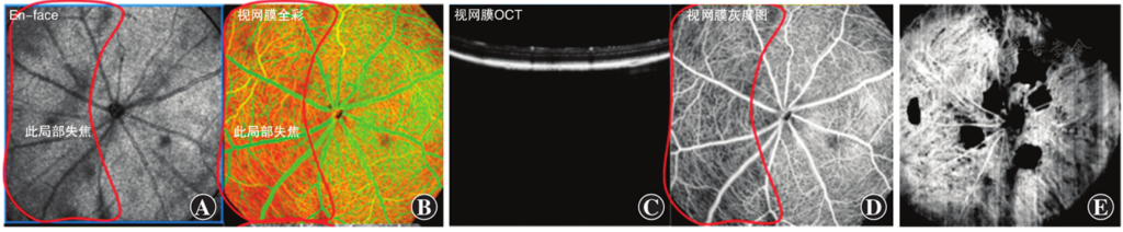
Figure 10 Low-quality OCTA map of retina and choroid A: En face image of local retinal defocus OCT B: Picture A corresponding to retinal OCTA map C: Picture A corresponding to retinal OCTA map D: Picture A corresponding to retinal OCTA gray scale E: Picture A corresponding to choroid OCTA map OCT: optical coherence tomography; OCTA: optical coherence tomography angiography
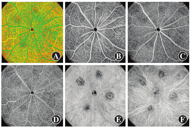
Firure 11 OCTA images of retina and choroid in laser-induced choroid neovascularization model in C57BL/6 mice A: Retinal color map B: Gray map of nerve fiber layer C: Gray map of inner plexiform layer D: Gray map of outer plexiform layer E: Gray map of superficial choroid F: Gray map of deep choroid OCTA: optical coherence tomography angiography
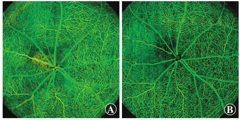
Figure 12 OCTA pictures at different time points after C57BL/6 mouse retinal vein occlusion model establishment A: Day 5 after modeling B: Day 12 after modeling OCTA: optical coherence tomography angiography
References click to collapse
[1] Huang D, Swanson EA, Lin CP, et al. Optical coherence tomography [J]. Science, 1991, 254 (5035):1178‑1181. DOI: 10.1126/science.1957169.
[2] Meinke J, Raming K, Kessler C, et al. High‑resolution optical coherence tomography correlates of Peau d’Orange in pseudoxanthoma elasticum [J]. JAMA Ophthalmol, 2026, 143 (12):1025‑1032. DOI: 10.1001/jamaophthalmol.2025.4031.
[3] Dauerman HL. Optical coherence tomography‑light and truth [J]. N Engl J Med, 2023, 389 (16):1523‑1525. DOI: 10.1056/NEJMe2309710.
[4] Gao SS, Jia Y, Zhang M, et al. Optical coherence tomography angiography [J]. Invest Ophthalmol Vis Sci, 2016, 57 (9):OCT27‑36. DOI: 10.1167/iovs.15‑19043.
[5] Zhang A, Zhang Q, Chen CL, et al. Methods and algorithms for optical coherence tomography‑based angiography: a review and comparison [J]. J Biomed Opt, 2015, 20 (10):100901. DOI: 10.1117/1.JBO.20.10.100901.
[6] Ang M, Tan A, Cheung C, et al. Optical coherence tomography angiography: a review of current and future clinical applications [J]. Graefes Arch Clin Exp Ophthalmol, 2018, 256 (2):237‑245. DOI: 10.1007/s00417‑017‑3896‑2.
[7] 徐桂花,宋跃。眼前节相干光断层扫描仪在眼科的应用 [J]. 国际眼科杂志,2008, 8 (10):2110‑2112.
Xu GH, Song Y. Assessment of ophthalmologic application with Visante OCT [J]. Int Eye Sci, 2008, 8 (10):2110‑2112.
[8] Kishi S. Impact of swept source optical coherence tomography on ophthalmology [J]. Taiwan J Ophthalmol, 2016, 6 (2):58‑68. DOI: 10.1016/j.tjo.2015.09.002.
[9] Podoleanu AG. Optical coherence tomography [J]. J Microsc, 2012, 247 (3):209‑219. DOI: 10.1111/j.1365‑2818.2012.03619.x.
[10] Puk O, de Angelis MH, Graw J. Longitudinal fundus and retinal studies with SD‑OCT: a comparison of five mouse inbred strains [J]. Mamm Genome, 2013, 24 (5‑6):198‑205. DOI: 10.1007/s00335‑013‑9457‑z.
[11] Bell BA, Kaul C, Dunaief JL, et al. A comparison of optophysiological biomarkers of photoreceptor stress and phototoxicity in BALB/cJ, B6 (Cg)‑Tyrc‑2J/J, and C57Bl/6J mouse strains [J]. Front Ophthalmol (Lausanne), 2023, 3:1128311. DOI: 10.3389/fopht.2023.1128311.
[12] Chauhan P, Kho AM, FitzGerald P, et al. Subcellular comparison of visible‑light optical coherence tomography and electron microscopy in the mouse outer retina [J]. Invest Ophthalmol Vis Sci, 2022, 63 (9):10. DOI: 10.1167/iovs.63.9.10.
[13] Ma Y, Dong R, Song T, et al. Evaluating the effects of three anesthetic regimens on retinal structure and function in the living mouse eye using a customized bimodal OCT/ORG system [J]. J Phys D: Appl Phys, 2025, 58 (3):035401. DOI: 10.1088/1361‑6463/ad89cc.
[14] Gandhi JK, Roy Chowdhury U, Manzar Z, et al. Differential intraocular pressure measurements by tonometry and direct cannulation after treatment with soluble adenylyl cyclase inhibitors [J]. J Ocul Pharmacol Ther, 2017, 33 (8):574‑581. DOI: 10.1089/jop.2017.0027.
[15] Rakymzhan A, Li Y, Tang P, et al. Differences in cerebral blood vasculature and flow in awake and anesthetized mouse cortex revealed by quantitative optical coherence tomography angiography [J]. J Neurosci Methods, 2021, 353:109094. DOI: 10.1016/j.jneumeth.2021.109094.
[16] Meleppat RK, Valente D, Lee S, et al. On‑axis full‑field swept‑source optical coherence tomography for murine retinal imaging [J]. Opt Lett, 2024, 49 (16):4630‑4633. DOI: 10.1364/OL.531116.
[17] Bian H, Wang J, Hong C, et al. GPU‑accelerated image registration algorithm in ophthalmic optical coherence tomography [J]. Biomed Opt Express, 2023, 14 (1):194‑207. DOI: 10.1364/BOE.479343.
[18] Podkowinski D, Sharian Varnousfaderani E, Simader C, et al. Impact of B‑scan averaging on spectralis optical coherence tomography image quality before and after cataract surgery [J]. J Ophthalmol, 2017, 2017:8148047. DOI: 10.1155/2017/8148047.
[19] Sakamoto A, Hangai M, Yoshimura N. Spectral‑domain optical coherence tomography with multiple B‑scan averaging for enhanced imaging of retinal diseases [J]. Ophthalmology, 2008, 115 (6):1071‑1078. DOI: 10.1016/j.ophtha.2007.09.001.
[20] 彭希,刘谊。角膜厚度的测量及其临床意义 [J]. 临床眼科杂志,2013, 21 (2):182‑187.
Peng X, Liu Y. Measurement of corneal thickness and its clinical significance [J]. J Clin Ophthalmol, 2013, 21 (2):182‑187.
[21] Meek KM, Knupp C. Corneal structure and transparency [J]. Prog Retin Eye Res, 2015, 49:1‑16. DOI: 10.1016/j.preteyeres.2015.07.001.
[22] Müller LJ, Marfurt CF, Kruse F, et al. Corneal nerves: structure, contents and function [J]. Exp Eye Res, 2003, 76 (5):521‑542. DOI: 10.1016/s0014‑4835 (03) 00050‑2.
[23] 郑广瑛,连元君,温成林,等。实验动物兔角膜 Orbscan‑Ⅱ 检测 [J]. 中华实验眼科杂志,2007, 25 (4):281‑283. DOI: 10.3760/cma.j.issn.2095‑0160.2007.04.011.
Zheng GY, Lian YJ, Wen CL, et al. Orbscan‑Ⅱ examination of cornea in normal rabbits [J]. Chin J Exp Ophthalmol, 2007, 25 (4):281‑283. DOI: 10.3760/cma.j.issn.2095‑0160.2007.04.011.
[24] 《眼轴长度在近视防控管理中的应用专家共识 (2023)》专家组。眼轴长度在近视防控管理中的应用专家共识 (2023)[J]. 中华实验眼科杂志,2024, 42 (1):1‑11. DOI: 10.3760/cma.j.cn115989‑20231010‑00121.
Expert Workgroup of Expert consensus on the application of axial length in myopia prevention and control management (2023). Expert consensus on the application of axial length in myopia prevention and control management (2023)[J]. Chin J Exp Ophthalmol, 2024, 42 (1):1‑11. DOI: 10.3760/cma.j.cn115989‑20231010‑00121.
[25] Kojima T, Tamaoki A, Yoshida N, et al. Evaluation of axial length measurement of the eye using partial coherence interferometry and ultrasound in cases of macular disease [J]. Ophthalmology, 2010, 117 (9):1750‑1754. DOI: 10.1016/j.ophtha.2010.01.018.
[26] Wolffsohn JS, Kollbaum PS, Berntsen DA, et al. IMI‑clinical myopia control trials and instrumentation report [J]. Invest Ophthalmol Vis Sci, 2019, 60 (3):M132‑M160. DOI: 10.1167/iovs.18‑25955.
[27] Brás J, Sickenberger W, Hirnschall N, et al. Cataract quantification using swept‑source optical coherence tomography [J]. J Cataract Refract Surg, 2018, 44 (12):1478‑1481. DOI: 10.1016/j.jcrs.2018.08.009.
[28] Huang J, Chen H, Li Y, et al. Comprehensive comparison of axial length measurement with three swept‑source OCT‑based biometers and partial coherence interferometry [J]. J Refract Surg, 2019, 35 (2):115‑120. DOI: 10.3928/1081597X‑20190109‑01.
[29] Grünert U, Martin PR. Cell types and cell circuits in human and non‑human primate retina [J]. Prog Retin Eye Res, 2020:100844. DOI: 10.1016/j.preteyeres.2020.100844.
[30] Clinical Laboratory Animal Medicine [M]. Welly Blackwell, 4th edition.
[31] Masland RH. The fundamental plan of the retina [J]. Nat Neurosci, 2001, 4 (9):877‑886. DOI: 10.1038/nn0901‑877.
[32] Hoon M, Okawa H, Della Santina L, et al. Functional architecture of the retina: development and disease [J]. Prog Retin Eye Res, 2014, 42:44‑84. DOI: 10.1016/j.preteyeres.2014.06.003.
[33] Liang X, Graf BW, Boppart SA. Imaging engineered tissues using structural and functional optical coherence tomography [J]. J Biophotonics, 2009, 2 (11):643‑655. DOI: 10.1002/jbio.200910048.
[34] Chen CL, Wang RK. Optical coherence tomography based angiography [Invited][J]. Biomed Opt Express, 2017, 8 (2):1056‑1082. DOI: 10.1016/j.boe.2016.12.027.
[35] 陈慧黎,梅立新,戴巧云,等。光学相干断层扫描血管成像在眼科的应用进展 [J]. 国际眼科杂志,2021, 21 (11):1918‑1921. DOI: 10.3980/j.issn.1672‑5123.2021.11.170.
Chen HL, Mei LX, Dai QY, et al. Application of optical coherence tomography angiography in ophthalmology [J]. Int Eye Sci, 2021, 21 (11):1918‑1921. DOI: 10.3980/j.issn.1672‑5123.2021.11.170.
[36] Koustenis A, Harris A, Gross J, et al. Optical coherence tomography angiography: an overview of the technology and an assessment of applications for clinical research [J]. Br J Ophthalmol, 2017, 101 (1):16‑20. DOI: 10.1136/bjophthalmol‑2016‑309389.
[37] Jia Y, Tan O, Tokayer J, et al. Split‑spectrum amplitude‑decorrelation angiography with optical coherence tomography [J]. Opt Express, 2012, 20 (4):4710‑4725. DOI: 10.1364/OE.20.004710.
[38] Freddo TF. Ultrastructure of the iris [J]. Microsc Res Tech, 1996, 33 (5):369‑389. DOI: 10.1002/(SICI) 1097‑0029 (19960401) 33:5<369::AID‑JEMT1>3.0.CO;2‑J.
[39] 沈锦琰,陈萱,龚学春,等。虹膜成像的研究进展 [J]. 临床眼科杂志,2023, 31 (2):174‑178. DOI: 10.3969/j.issn.1006‑8422.2023.02.020.
Shen JY, Chen X, Gong XC, et al. Research progress in iris imaging [J]. J Clin Ophthalmol, 2023, 31 (2):174‑178. DOI: 10.3969/j.issn.1006‑8422.2023.02.020.
[40] Pan WW, Wubben TJ, Besirli CG. Photoreceptor metabolic reprogramming: current understanding and therapeutic implications [J]. Commun Biol, 2021, 4 (1):245. DOI: 10.1038/s42003‑021‑01765‑3.
[41] Fu Z, Sun Y, Cakir B, et al. Targeting neurovascular interaction in retinal disorders [J]. Int J Mol Sci, 2020, 21 (4):503. DOI: 10.3390/ijms21041503.
[42] Usui Y, Westenskow PD, Kurihara T, et al. Neurovascular crosstalk between interneurons and capillaries is required for vision [J]. J Clin Invest, 2015, 125 (6):2335‑2346. DOI: 10.1172/JCI80297.
[43] Bharadwaj AS, Appukuttan B, Wilmarth PA, et al. Role of the retinal vascular endothelial cell in ocular disease [J]. Prog Retin Eye Res, 2013, 32:102‑180. DOI: 10.1016/j.preteyeres.2012.08.004.
[44] Bek T. Regional morphology and pathophysiology of retinal vascular disease [J]. Prog Retin Eye Res, 2013, 36:247‑259. DOI: 10.1016/j.preteyeres.2013.07.002.
[45] Nickla DL, Wallman J. The multifunctional choroid [J]. Prog Retin Eye Res, 2010, 29 (2):144‑168. DOI: 10.1016/j.preteyeres.2009.12.002.
[46] 黄晴予,武志峰。脉络膜结构与功能的研究进展 [J]. 临床眼科杂志,2023, 31 (1):83‑86. DOI: 10.3969/j.issn.1006‑8422.2023.01.022.
Huang QY, Wu ZF. Research progress on the structure and function of the choroid [J]. J Clin Ophthalmol, 2023, 31 (1):83‑86. DOI: 10.3969/j.issn.1006‑8422.2023.01.022.
[47] Bermudez MA, Vicente AF, Romero MC, et al. Time course of cold cataract development in anesthetized mice [J]. Curr Eye Res, 2011, 36 (3):278‑284. DOI: 10.3109/02713683.2010.542868.
[48] Liu X, Wang CH, Dai C, et al. Effect of contact lens on optical coherence tomography imaging of rodent retina [J]. Curr Eye Res, 2013, 38 (12):1235‑1240. DOI: 10.3109/02713683.2013.815218.
[49] 詹红微,邱泽文,李慧玲,等。三种麻醉药物对小鼠麻醉效果的比较 [J]. 实验动物科学,2012, 29 (4):19‑24.
Zhan HW, Qiu ZW, Li HL, et al. Anesthetic effects of three kind of narcotic drugs on mice [J]. Lab Anim Sci, 2012, 29 (4):19‑24.