Authors:Sun Xiuli, Gu Yixuan, Zhu Lei,Tian Lei, Wang Yinghui,Jie Ying
Corresponding author:Jie Ying, Email:jie_yingcn@aliyun.com
Published:2026-08-10
DOI:10.3760/cma.j.cn115989-20250916-00300
ABSTRACT
Objective To evaluate the accuracy and repeatability of fluorescence PCR in detecting TGFBI gene mutation in patients with corneal dystrophy.
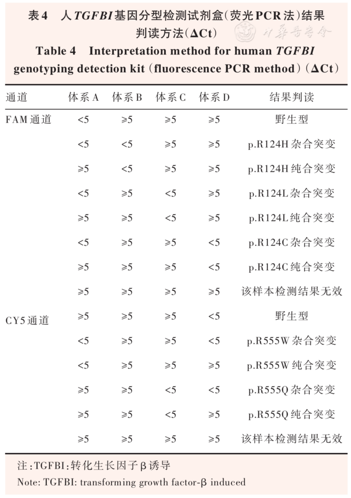
Methods ARMS-PCR combined with TaqMan probe method was used for genotyping the R124H, R124C, R555W, and R555Q loci (covering heterozygous and homozygous mutation) in the TGFBI gene in peripheral blood samples from 240 patients with corneal dystrophy who were treated at Beijing Tongren Hospital, Capital Medical University from January 2021 to August 2025, and the results were compared with Sanger sequencing results. The intra-batch and inter-batch test results of fluorescence PCR were analyzed to evaluate their repeatability. The association between genotypes and clinical phenotypes was analyzed. This study adhered to the principles of the Declaration of Helsinki. The study protocol was approved by the Ethics Committee of Beijing Tongren Hospital, Capital Medical University (No. TREC2023012), and written informed consent was obtained from all patients.
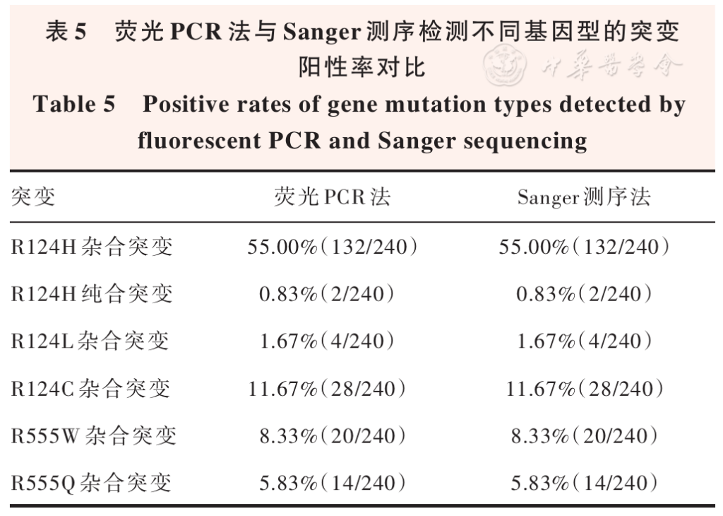
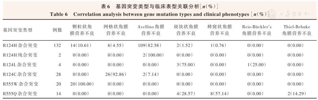
Results The positive rate of pathogenic gene mutations detected by the human TGFBI gene mutation detection kit (fluorescence PCR method) and Sanger sequencing in 240 samples was 83.33%(200/240). The positive rates of R124H heterozygous mutation, R124H homozygous mutation, R124L heterozygous mutation, R124C heterozygous mutation, R555W heterozygous mutation and R555Q heterozygous mutation were 55.00%(132/240), 0.83%(2/240), 1.67%(4/240), 11.67%(28/240), 8.33%(20/240), 5.83%(14/240), respectively. The total positive rate was 83.33%(200/240), and the wild-type detection rate was 16.67%(40/240). The total coincidence rate of the two methods was 100.00%. The repeated results of intra-batch and inter-batch for TGFBI gene mutation detected by fluorescence PCR were completely consistent. Analysis of genotypes and clinical phenotypes in 200 patients with positive gene mutations showed that the differences in the distribution of clinical phenotypes among patients with different genotypes was statistically significant (χ2=498.474, P<0.001). Cramer V=0.706.
Conclusions Fluorescence PCR is highly accurate and reproducible in detecting TGFBI gene hot mutation, which is suitable for detecting gene mutation in patients with corneal dystrophy in clinical practice.
KEYWORDS:
Corneal dystrophy;TGFBI gene ;Fluorescence PCR;Sanger sequencing;Accuracy;Repeatability
COPYRIGHTS:
Copyright by Chinese Medical Association
No content published by the journals of Chinese Medical Association may be reproduced or abridged without authorization. Please do not use or copy the layout and design of the journals without permission.
All articles published represent the opinions of the authors, and do not reflect the official policy of the Chinese Medical Association or the Editorial Board, unless this is clearly specified.
Authors Info & Affiliations
Sun Xiuli
Beijing Institute of Ophthalmology, Beijing Tongren Eye Center, Beijing Tongren Hospital, Capital Medical University, Beijing Key Laboratory of Ophthalmology and Visual Science, Beijing 100730, China
Gu Yixuan
Beijing Institute of Ophthalmology, Beijing Tongren Eye Center, Beijing Tongren Hospital, Capital Medical University, Beijing Key Laboratory of Ophthalmology and Visual Science, Beijing 100730, China
Zhu Lei
Beijing Institute of Ophthalmology, Beijing Tongren Eye Center, Beijing Tongren Hospital, Capital Medical University, Beijing Key Laboratory of Ophthalmology and Visual Science, Beijing 100730, China
Tian Lei
Beijing Institute of Ophthalmology, Beijing Tongren Eye Center, Beijing Tongren Hospital, Capital Medical University, Beijing Key Laboratory of Ophthalmology and Visual Science, Beijing 100730, China
Wang Yinghui
Beijing Institute of Ophthalmology, Beijing Tongren Eye Center, Beijing Tongren Hospital, Capital Medical University, Beijing Key Laboratory of Ophthalmology and Visual Science, Beijing 100730, China
Jie Ying
Beijing Institute of Ophthalmology, Beijing Tongren Eye Center, Beijing Tongren Hospital, Capital Medical University, Beijing Key Laboratory of Ophthalmology and Visual Science, Beijing 100730, China
Figures & Tables
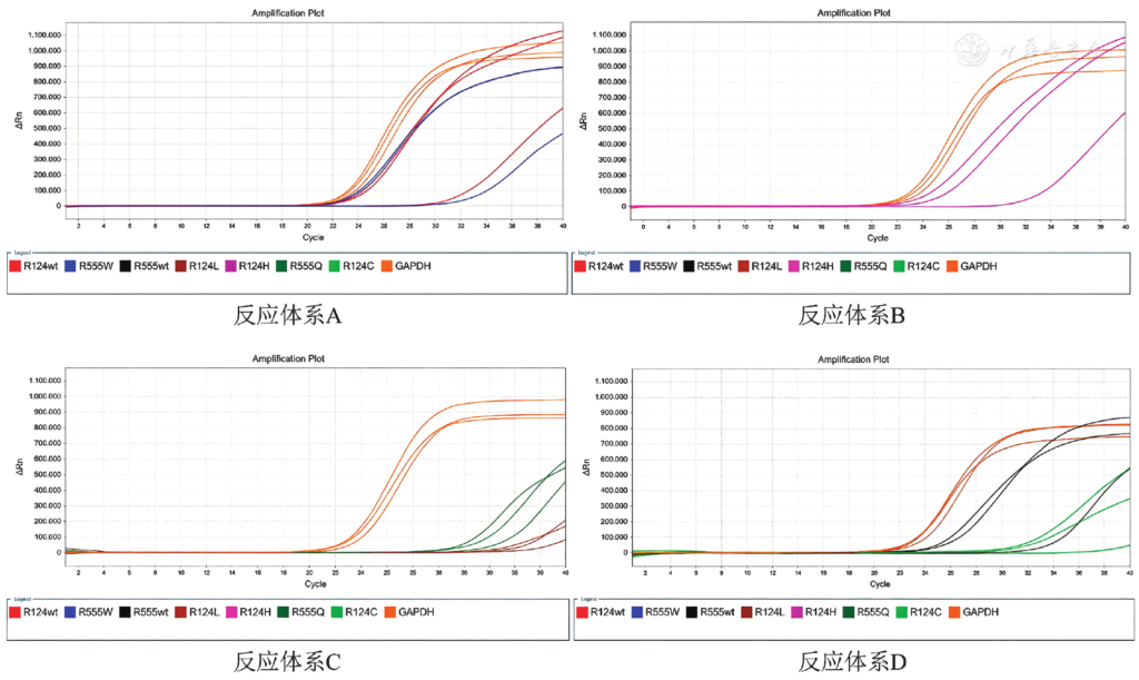
Figure 1 Amplification curve of reference substance wt: wild type; GAPDH: glyceraldehyde-3-phos phate dehydrogenase
References click to collapse
[1] Li T, Wu S, Wen Y, et al. Mutation analysis of the TGFBI gene in pedigrees of lattice corneal dystrophy in Eastern China [J]. Ophthalmic Genet, 2022, 43 (5):594‑601. DOI: 10.1080/13816810.2022.2068616.
[2] Sciriha GG, Sultana J, Borg J. Identifying and categorizing compounds that reduce corneal transforming growth factor beta induced protein levels: a scoping review [J]. Expert Rev Clin Pharmacol, 2022, 15 (12):1423‑1442. DOI: 10.1080/17512433.2022.2142560.
[3] Han SB, Anandalakshmi V, Wong CW, et al. Genotypic homogeneity in distinctive transforming growth factor‑beta induced (TGFBI) protein phenotypes [J]. Int J Mol Sci, 2021, 22 (3):1230. DOI: 10.3390/ijms22031230.
[4] Munier FL, Korvatska E, Djemaï A, et al. Kerato‑epithelin mutations in four 5q31‑linked corneal dystrophies [J]. Nat Genet, 1997, 15 (3):247‑251. DOI: 10.1038/ng0397‑247.
[5] Lisch W, Weiss JS. Early and late clinical landmarks of corneal dystrophies [J]. Exp Eye Res, 2020, 198:108139. DOI: 10.1016/j.exer.2020.108139.
[6] Han KE, Choi SI, Kim TI, et al. Pathogenesis and treatments of TGFBI corneal dystrophies [J]. Prog Retin Eye Res, 2016, 50:67‑88. DOI: 10.1016/j.preteyeres.2015.11.002.
[7] Cui X, Shu L, Liu B, et al. Rare TGFBI mutation c.1553T>G p.(L518R) in lattice corneal dystrophy: comprehensive clinical and genetic analysis in a Chinese family [J]. Am J Ophthalmol, 2025, 279:127‑136. DOI: 10.1016/j.ajo.2025.07.012.
[8] Zacharogianni M, Papandreou NC, Marinakis NM, et al. Clinical and structural characterization of a novel TGFBI mutation linked to a lattice corneal dystrophy variant in a Greek family [J]. Am J Ophthalmol, 2025, 274:112‑121. DOI: 10.1016/j.ajo.2025.03.003.
[9] Weiss JS, Willoughby CE, Abad‑Morales V, et al. Update on the corneal dystrophies‑genetic testing and therapy [J]. Cornea, 2022, 41 (11):1337‑1344. DOI: 10.1097/ICO.0000000000002857.
[10] Skonier J, Neubauer M, Madisen L, et al. cDNA cloning and sequence analysis of beta ig‑h3, a novel gene induced in a human adenocarcinoma cell line after treatment with transforming growth factor‑beta [J]. DNA Cell Biol, 1992, 11 (7):511‑522. DOI: 10.1089/dna.1992.11.511.
[11] Sciriha GG, Borg J, Sultana J, et al. Transcriptome analysis of TGFBI knockdown vs normal corneal epithelial cells: implications for TGFBI corneal dystrophy treatment [J]. Biochem Genet, 2026, 64 (3):3451‑3480. DOI: 10.1007/s10528‑025‑11191‑3.
[12] Li F, He J, Bai H, et al. An Arg124Cys mutation in transforming growth factor β‑induced gene associated with lattice corneal dystrophy type I in a Chinese pedigree [J]. Indian J Ophthalmol, 2022, 70 (1):85‑89. DOI: 10.4103/ijo.IJO_33_21.
[13] Tan J, Zhang X, Wei X, et al. Application of the TaqMan ARMS‑PCR approach for genotyping drug‑induced hearing loss using dried blood samples [J]. Curr Issues Mol Biol, 2024, 46 (6):5454‑5466. DOI: 10.3390/cimb46060326.
[14] 许可,孙旭光,谢玥,等。角膜营养不良患者 TGFBI 基因突变特点及基因型‑表型关系 [J]. 中华眼视光学与视觉科学杂志,2016, 18 (3):153‑159. DOI: 10.3760/cma.j.issn.1674‑845X.2016.03.006.
Xu K, Sun XG, Xie Y, et al. Gene mutation and genotype‑phenotype description in corneal dystrophy associated with TGFBI genes [J]. Chin J Optom Ophthalmol Vis Sci, 2016, 18 (3):153‑159. DOI: 10.3760/cma.j.issn.1674‑845X.2016.03.006.