Authors:Gao Linghan, Duan Wenxuan, Duan Yanli, Liu Yang
Corresponding author: Liu Yang, Email:ldyy_liuyang@163.com
Published:2026-10-10
DOI:10.3760/cma.j.cn115989-20260310-00112
ABSTRACT
This report describes the clinical features, diagnostic workup, management, and short-term follow-up of a patient with clinically diagnosed xeroderma pigmentosum (XP)-related keratoconjunctivitis. Slit-lamp examination and anterior segment photography demonstrated premature aging and leathery changes of the bilateral eyelid skin, lower eyelid ectropion, and indistinct meibomian gland orifices, indicating potential involvement of the lid margin and meibomian glands. Additional findings included conjunctival mixed hyperemia, corneal neovascularization, corneal ulcerative infiltration, and stromal edema. In view of the characteristic cutaneous lesions on sun-exposed areas, a history of parental consanguinity, and the above ocular surface abnormalities, XP was diagnosed clinically, and the ocular manifestations were attributed to XP-associated keratoconjunctivitis. Genetic testing, skin biopsy, and supplementary ocular examinations (including corneal scraping and culture, corneal sensation assessment, fluorescein staining score, tear break-up time, and Schirmer test) were not performed at the patient’s request. Therefore, the diagnosis in this case was established primarily on clinical phenotyping. The treatment regimen consisted of lid hygiene, warm compresses, topical antimicrobial agents, ocular lubrication, and short-term anti-inflammatory therapy. The patient was also counseled on strict ultraviolet protection and scheduled for long-term follow-up. One week after treatment, repeat anterior segment photography revealed alleviated lid margin hyperemia, reduced area of corneal ulcerative infiltration, and less hyperemia of corneal neovascularization. This case suggests that XP-related ocular surface disease should be suspected in patients with refractory bilateral keratoconjunctivitis who present with premature eyelid skin aging, madarosis, lower lid ectropion, pigmentary abnormalities of sun-exposed skin, and a family history of consanguinity.
KEYWORDS:
Xeroderma pigmentosum; Keratoconjunctivitis; Ocular surface disease; Corneal neovascularization; Case report
COPYRIGHTS:
Copyright by Chinese Medical Association
No content published by the journals of Chinese Medical Association may be reproduced or abridged without authorization. Please do not use or copy the layout and design of the journals without permission.
All articles published represent the opinions of the authors, and do not reflect the official policy of the Chinese Medical Association or the Editorial Board, unless this is clearly specified.
Authors Info & Affiliations
Gao Linghan
The First Clinical Medical College of Lanzhou University, Lanzhou 730000, China
Duan Wenxuan
The First Clinical Medical College of Lanzhou University, Lanzhou 730000, China
Duan Yanli
The First Clinical Medical College of Lanzhou University, Lanzhou 730000, China
Liu Yang
Ophthalmology Department of Lanzhou University First Hospital, Lanzhou 730000, China
Figures & Tables
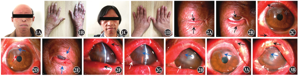
Figure 1 Facial and hand photographs of the patient and her elder sister A, B: The patient presented with dense freckle-like pigmentation on sun-exposed areas including the face, hands and upper arms. Pigmented and depigmented areas were intermingled, accompanied by prominent telangiectasia, rough and thickened skin, and premature aging changes. C, D: The patient’s elder sister exhibited similar cutaneous lesions on the face and hands, yet with milder severity.
Figure 2 Anterior segment photographs of both eyes before treatment A: Dry, leathery changes of the right eyelid skin (black arrow). B: Right lower eyelid ectropion with extensive madarosis (black arrow). C: Hypertrophy and hyperemia of the right upper and lower lid margins, with indistinct meibomian gland orifices (black arrow). D: Mixed hyperemia of the right bulbar conjunctiva (white arrow); an approximately 2-mm ulcerative infiltrate was noted superiorly on the cornea (red arrow), with corneal neovascularization extending about 2 mm from the limbus (blue arrow). E: Dry, leathery changes of the left eyelid skin (blue arrow), lower eyelid ectropion and extensive madarosis (red arrow). F: Hypertrophy and hyperemia of the left upper and lower lid margins, with indistinct meibomian gland orifices (black arrow). G: Mixed hyperemia of the left bulbar conjunctiva (white arrow); an approximately 5-mm ulcerative infiltrate was present in the pupillary area of the cornea, with focal scar formation (blue arrow). H: Temporoinferior neovascularization extending approximately 3 mm from the left corneal limbus (white arrow); superior neovascularization extended into the corneal pupillary zone (blue arrow), associated with corneal stromal edema.
Figure 3 Anterior segment photographs of both eyes one week after treatment A: Decreased hyperemia of the right lid margin (black arrow), and reduced size of the corneal ulcerative infiltrate (blue arrow). B: Decreased hyperemia of the left lid margin (black arrow), attenuated hyperemia of corneal neovascularization (white arrow), and reduced size of the corneal ulcerative infiltrate (blue arrow).
▼click to see the rest
References
[1] Lehmann AR, McGibbon D, Stefanini M. Xeroderma pigmentosum[J]. Orphanet J Rare Dis, 2011, 6∶70. DOI: 10.1186/1750‐1172‐6‐70.
▼click to see more references
[2] Leung AK, Barankin B, Lam JM, et al. Xeroderma pigmentosum: an updated review[J]. Drugs Context, 2022, 11:2022-2-5. DOI: 10.7573/dic.2022-2-5.
[3] Brooks BP, Thompson AH, Bishop RJ, et al. Ocular manifestations of xeroderma pigmentosum: long-term follow-up highlights the role of DNA repair in protection from sun damage[J]. Ophthalmology, 2013, 120(7):1324-1336. DOI: 10.1016/j.ophtha.2012.12.044.
[4] Kraemer KH, Lee MM, Scotto J. Xeroderma pigmentosum. Cutaneous, ocular, and neurologic abnormalities in 830 published cases[J]. Arch Dermatol, 1987, 123(2):241-250. DOI: 10.1001/archderm.123.2.241.
[5] Suarez MJ, Rivera-Michlig R, Dubovy S, et al. Clinicopathological features of ophthalmic neoplasms arising in the setting of xeroderma pigmentosum[J]. Ocul Oncol Pathol, 2015, 2(2):112-121. DOI: 10.1159/000441527.
[6] Ramkumar HL, Brooks BP, Cao X, et al. Ophthalmic manifestations and histopathology of xeroderma pigmentosum: two clinicopathological cases and a review of the literature[J]. Surv Ophthalmol, 2011, 56(4):348-361. DOI: 10.1016/j.survophthal.2011.03.001.
[7] Marcos A, Freitas D, Hazarbassanov RM, et al. Evaluation of meibomian gland dysfunction using meibography in patients with xeroderma pigmentosum[J]. Arq Bras Oftalmol, 2024, 87(2):e20220319. DOI: 10.5935/0004-2749.2022-0319.
[8] Sarkany R, Canfield M, Morgan M, et al. Ultraviolet radiation exposure to the face in patients with xeroderma pigmentosum and healthy controls: applying a novel methodology to define photoprotection behaviour[J]. Br J Dermatol, 2022, 186(4):713-720. DOI: 10.1111/bjd.20899.
[9] 柯丹丹, 刘荣, 罗班, 等. 迟发性着色性干皮病继发两种眼睑恶性肿瘤1例[J]. 中华实验眼科杂志, 2020, 38(9):765-766. DOI: 10.3760/cma.j.cn115989-20200825-00609. Ke DD, Liu R, Luo B, et al. A case of tardive xeroderma pigmentosum secondary with two kinds of eyelid malignant tumors[J]. Chin J Exp Ophthalmol, 2020, 38(9):765-766. DOI: 10.3760/cma.j.cn115989-20200825-00609.
[10] Malikireddy S. Xeroderma pigmentosum with ocular surface squamous neoplasia: a case report[J]. Cureus, 2024, 16(1):e53204. DOI: 10.7759/cureus.53204.
[11] Wang Y, Cai L, Feng H, et al. Treatment of corneoscleral mixed hemangioma by intrastromal lenticule transplantation in a case of xeroderma pigmentosum: a case report[J]. BMC Ophthalmol, 2025, 25(1):277. DOI: 10.1186/s12886-025-04087-8.
[12] Rajagopal R, Srinivasan B, Iyer G, et al. Management of coexisting bilateral ocular surface disease in xeroderma pigmentosum-sequence and outcomes[J]. Eur J Ophthalmol, 2023, 33(4):NP9-NP12. DOI: 10.1177/11206721221100903.