Authors:Wang Junwen, Ma Yunxi, Sun Xufang
Corresponding author: Sun Xufang, Email: sunxufang2016@163.com
Published:2026-09-10
DOI: Neovascular age-related macular degeneration (nAMD) is one of the leading causes of central vision loss and irreversible blindness in the elderly population. Its core pathological features include choroidal neovascularization, exudation, hemorrhage, and scarring. In recent years, although anti-vascular endothelial growth factor (anti-VEGF) therapy has significantly improved short-term visual outcomes in nAMD patients, approximately 30% of patients still experience suboptimal response, frequent recurrence, and long-term progression to geographic atrophy and subretinal fibrosis in clinical practice. These limitations suggest that, in addition to the VEGF pathway, other critical pathogenic mechanisms remain to be elucidated. Multi-omics and translational medicine studies have demonstrated that aberrant activation of the complement system plays a pivotal role throughout the initiation and progression of nAMD. Complement cascade activation not only directly mediates retinal pigment epithelial cell injury, disruption of Bruch membrane homeostasis, and recruitment of inflammatory cells, but also accelerates CNV formation and disease progression by modulating the immune microenvironment, promoting VEGF release, and driving fibrotic remodeling. Currently, complement targets such as C3, C5, CFB, and CFD have become major hotspots in AMD therapeutic research. Various complement inhibitors have shown promising potential in clinical trials, driving the development of dual-target or multi-target synergistic strategies, particularly “complement inhibition combined with anti-VEGF” therapy. This article systematically integrates the latest advances in genetics, histopathology, multimodal imaging, and biomarkers to deeply analyze the pathogenic mechanisms of the complement system in nAMD and its association with treatment resistance and compensatory responses. It also tracks the clinical translation of multi-target combination strategies, with the aim of providing a forward-looking translational medicine perspective for advancing nAMD management from single anti-angiogenic therapy toward multi-mechanism synergistic and precision individualized treatment.
ABSTRACT
Neovascular age-related macular degeneration (nAMD) is one of the leading causes of central vision loss and irreversible blindness in the elderly population. Its core pathological features include choroidal neovascularization, exudation, hemorrhage, and scarring. In recent years, although anti-vascular endothelial growth factor (anti-VEGF) therapy has significantly improved short-term visual outcomes in nAMD patients, approximately 30% of patients still experience suboptimal response, frequent recurrence, and long-term progression to geographic atrophy and subretinal fibrosis in clinical practice. These limitations suggest that, in addition to the VEGF pathway, other critical pathogenic mechanisms remain to be elucidated. Multi-omics and translational medicine studies have demonstrated that aberrant activation of the complement system plays a pivotal role throughout the initiation and progression of nAMD. Complement cascade activation not only directly mediates retinal pigment epithelial cell injury, disruption of Bruch membrane homeostasis, and recruitment of inflammatory cells, but also accelerates CNV formation and disease progression by modulating the immune microenvironment, promoting VEGF release, and driving fibrotic remodeling. Currently, complement targets such as C3, C5, CFB, and CFD have become major hotspots in AMD therapeutic research. Various complement inhibitors have shown promising potential in clinical trials, driving the development of dual-target or multi-target synergistic strategies, particularly “complement inhibition combined with anti-VEGF” therapy. This article systematically integrates the latest advances in genetics, histopathology, multimodal imaging, and biomarkers to deeply analyze the pathogenic mechanisms of the complement system in nAMD and its association with treatment resistance and compensatory responses. It also tracks the clinical translation of multi-target combination strategies, with the aim of providing a forward-looking translational medicine perspective for advancing nAMD management from single anti-angiogenic therapy toward multi-mechanism synergistic and precision individualized treatment.
KEYWORDS:
Age-related macular degeneration;Choroidal neovascularization;Complement;Clinical translation
COPYRIGHTS:
Copyright by Chinese Medical Association
No content published by the journals of Chinese Medical Association may be reproduced or abridged without authorization. Please do not use or copy the layout and design of the journals without permission.
All articles published represent the opinions of the authors, and do not reflect the official policy of the Chinese Medical Association or the Editorial Board, unless this is clearly specified.
Authors Info & Affiliations
Wang Junwen
Department of Ophthalmology, Tongji Hospital, Tongji Medical College, Huazhong University of Science and Technology, Hubei Key Laboratory of Otolaryngologic and Ophthalmic Disease (HUST), Wuhan 430030, China
Ma Yunxi
Department of Ophthalmology, Tongji Hospital, Tongji Medical College, Huazhong University of Science and Technology, Hubei Key Laboratory of Otolaryngologic and Ophthalmic Disease (HUST), Wuhan 430030, China
Sun Xufang
Department of Ophthalmology, Tongji Hospital, Tongji Medical College, Huazhong University of Science and Technology, Hubei Key Laboratory of Otolaryngologic and Ophthalmic Disease (HUST), Wuhan 430030, China
Figures & Tables
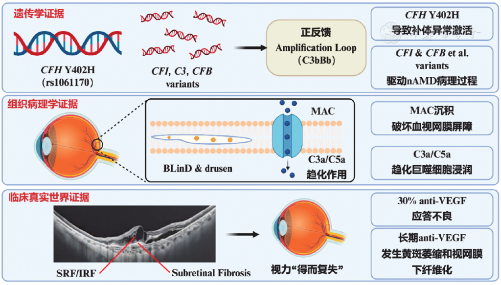
Figure 1 Role of abnormal complement activation in the occurrence and development of nAMD CFH: complement factor H;BLinD: basal linear deposits;MAC: membrane attack complex;SRF: subretinal fluid;IRF: intraretinal fluid;VEGF: vascular endothelial growth factor;nAMD: neovascular age-related macular degeneration
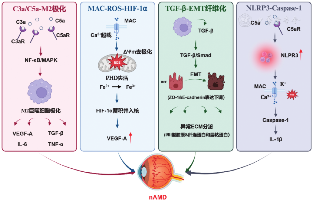
Figure 2 Mechanism of abnormal activation of complement promoting nAMD NF: nuclear factor;MAPK: mitogen-activated protein kinase;VEGF: vascular endothelial growth factor;MAC: membrane attack complex;ROS: reactive oxygen species;HIF: hypoxia-inducible factor;PHD: proline hydroxylase;TGF: transforming growth factor;RPE: retinal pigment epithelium;EMT: epithelial-mesenchymal transformation;ZO: tight junction protein;ECM: extracellular matrix; NLRP3: nucleotide binding oligomerization domain-like receptor protein 3;Caspase: Aspartine-specific cysteine protease;IL: interleukin;nAMD: neovascular age-related macular degeneration
References click to collapse
[1] Wu J, Shan S, Zhou J, et al. National, regional, and provincial prevalence of age-related macular degeneration in China in 2020: an updated systematic review and modelling study[J]. J Glob Health, 2026, 16:04062. DOI: 10.7189/jogh.16.04062.
[2] Flaxel CJ, Adelman RA, Bailey ST, et al. Age-related macular degeneration preferred practice pattern®[J]. Ophthalmology, 2020, 127(1):P1-P65. DOI: 10.1016/j.ophtha.2019.09.024.
[3] Ferris FL, Fine SL, Hyman L. Age-related macular degeneration and blindness due to neovascular maculopathy[J]. Arch Ophthalmol, 1984, 102(11):1640-1642. DOI: 10.1001/archopht.1984.01040031330019.
[4] Sorenson CM, Gurel Z, Song YS, et al. Thrombospondin-1, BIM and CFH polymorphisms and response to anti-VEGF treatment in neovascular age-related macular degeneration patients[J/OL]. PLoS One, 2024, 19(2):e0297135[2026-04-05]. https://pubmed.ncbi.nlm.nih.gov/38408093/. DOI: 10.1371/journal.pone.0297135.
[5] Comparison of Age-related Macular Degeneration Treatments Trials (CATT) Research Group, Maguire MG, Martin DF, et al. Five-year outcomes with anti-vascular endothelial growth factor treatment of neovascular age-related macular degeneration: the comparison of age-related macular degeneration treatments trials[J]. Ophthalmology, 2016, 123(8): 1751-1761.DOI: 10.1016/j.ophtha.2016.03.045.
[6] Mantel I, Dirani A, Zola M, et al. Macular atrophy incidence in anti-vascular endothelial growth factor-treated neovascular age-related macular degeneration: risk factor evaluation for individualized treatment need of ranibizumab or aflibercept according to an observe-and-plan regimen[J]. Retina,2019,39(5):906-917.DOI: 10.1097/IAE.0000000000002054.
[7] Sadda SR, Tuomi LL, Ding B, et al. Macular atrophy in the HARBOR Study for neovascular age-related macular degeneration[J]. Ophthalmology, 2018,125(6):878-886.DOI: 10.1016/j.ophtha.2017.12.026.
[8] Stewart C, Bhangu JS, Swarnkar PK, et al. Real-world outcomes of switching to faricimab in treatment-experienced and resistant neovascular age-related macular degeneration: a single-centre retrospective study[J]. Clin Ophthalmol,2026,20:591029.DOI: 10.2147/OPTH.S591029.
[9] Jia H, Lu B, Yuan Y, et al. A randomized, controlled trial of treat-and-extend vs. pro re nata regimen for neovascular age-related macular degeneration[J]. Front Med (Lausanne), 2022, 9:852519. DOI: 10.3389/fmed.2022.852519.
[10] Haines JL, Hauser MA, Schmidt S, et al. Complement factor H variant increases the risk of age-related macular degeneration[J]. Science, 2005, 308(5720):419-421. DOI: 10.1126/science.1110359.
[11] Edwards AO, Ritter R, Abel KJ, et al. Complement factor H polymorphism and age-related macular degeneration[J]. Science, 2005, 308(5720):421-424. DOI: 10.1126/science.1110189.
[12] Klein RJ, Zeiss C, Chew EY, et al. Complement factor H polymorphism in age-related macular degeneration[J]. Science, 2005, 308(5720): 385-389. DOI: 10.1126/science.1109557.
[13] Zipfel PF, Skerka C. Complement regulators and inhibitory proteins[J]. Nat Rev Immunol, 2009, 9(10):729-740. DOI: 10.1038/nri2620.
[14] Laine M, Jarva H, Seitsonen S, et al. Y402H polymorphism of complement factor H affects binding affinity to C-reactive protein[J]. J Immunol, 2007, 178(6):3831-3836. DOI: 10.4049/jimmunol.178.6.3831.
[15] 程心璇, 刘祖国, 廖怿. 补体异常活化促进年龄相关性黄斑变性发生的作用和机制[J]. 中华实验眼科杂志,2022, 40(12):1186-1191. DOI: 10.3760/cma.j.cn115989-20191023-00461. Cheng XX, Liu ZG, Liao Y. Role of complement dysregulation in age-related macular degeneration[J]. Chin J Exp Ophthalmol, 2022, 40(12):1186-1191. DOI: 10.3760/cma.j.cn115989-20191023-00461.
[16] Seddon JM, Yu Y, Miller EC, et al. Rare variants in CFI, C3 and C9 are associated with high risk of advanced age-related macular degeneration[J]. Nat Genet, 2013, 45(11):1366-1370. DOI: 10.1038/ng.2741.
[17] Gold B, Merriam JE, Zernant J, et al. Variation in factor B (BF) and complement component 2 (C2) genes is associated with age-related macular degeneration[J]. Nat Genet, 2006, 38(4):458-462. DOI: 10.1038/ng1750.
[18] Yates JR, Sepp T, Matharu BK, et al. Complement C3 variant and the risk of age-related macular degeneration[J]. N Engl J Med, 2007, 357(6): 553-561. DOI: 10.1056/NEJMoa072618.
[19] Armento A, Ueffing M, Clark SJ. The complement system in age-related macular degeneration[J]. Cell Mol Life Sci, 2021, 78(10):4487-4505. DOI: 10.1007/s00018-021-03796-9.
[20] Llorián-Salvador M, Byrne EM, Szczepan M, et al. Complement activation contributes to subretinal fibrosis through the induction of epithelial-to-mesenchymal transition (EMT) in retinal pigment epithelial cells[J]. J Neuroinflammation, 2022, 19(1):182. DOI: 10.1186/s12974-022-02546-3.
[21] Zareparsi S, Branham KE, Li M, et al. Strong association of the Y402H variant in complement factor H at 1q32 with susceptibility to age-related macular degeneration[J]. Am J Hum Genet, 2005, 77(1):149-153. DOI: 10.1086/431426.
[22] Fritsche LG, Igl W, Bailey JN, et al. A large genome-wide association study of age-related macular degeneration highlights contributions of rare and common variants[J]. Nat Genet, 2016, 48(2):134-143. DOI: 10.1038/ng.3448.
[23] Mullins RF, Russell SR, Anderson DH, et al. Drusen associated with aging and age-related macular degeneration contain proteins common to extracellular deposits associated with atherosclerosis, elastosis, amyloidosis, and dense deposit disease[J]. FASEB J, 2000, 14(7): 835-846.
[24] Bora PS, Sohn JH, Cruz JM, et al. Role of complement and complement membrane attack complex in laser-induced choroidal neovascularization[J]. J Immunol, 2005, 174(1):491-497. DOI: 10.4049/jimmunol.174.1.491.
[25] Joseph K, Kulik L, Coughlin B, et al. Oxidative stress sensitizes retinal pigmented epithelial (RPE) cells to complement-mediated injury in a natural antibody-, lectin pathway-, and phospholipid epitope-dependent manner[J]. J Biol Chem, 2013, 288(18):12753-12765. DOI: 10.1074/jbc.M112.421891.
[26] Curcio CA. Soft drusen in age-related macular degeneration: biology and targeting via the oil spill strategies[J]. Invest Ophthalmol Vis Sci, 2018, 59(4):AMD160-AMD181. DOI: 10.1167/iovs.18-24882.
[27] Kauppinen A, Paterno JJ, Blasiak J, et al. Inflammation and its role in age-related macular degeneration[J]. Cell Mol Life Sci, 2016, 73(9): 1765-1786. DOI: 10.1007/s00018-016-2147-8.
[28] Klaassen I, Van Noorden CJ, Schlingemann RO. Molecular basis of the inner blood-retinal barrier and its breakdown in diabetic macular edema and other pathological conditions[J]. Prog Retin Eye Res, 2013, 34:19-48. DOI: 10.1016/j.preteyeres.2013.02.001.
[29] Zhang J, Sheng X, Ding Q, et al. Subretinal fibrosis secondary to neovascular age-related macular degeneration: mechanisms and potential therapeutic targets[J]. Neural Regen Res, 2025, 20(2): 378-393. DOI: 10.4103/NRR.NRR-D-23-01642.
[30] Nozaki M, Raisler BJ, Sakurai E, et al. Drusen complement components C3a and C5a promote choroidal neovascularization[J]. Proc Natl Acad Sci U S A, 2006, 103(7):2328-2333. DOI: 10.1073/pnas.0408835103.
[31] Krause TA, Alex AF, Engel DR, et al. VEGF-production by CCR2-dependent macrophages contributes to laser-induced choroidal neovascularization[J/OL]. PLoS One, 2014, 9(4):e94313[2026-04-10]. https://pubmed.ncbi.nlm.nih.gov/24714223/. DOI: 10.1371/journal.pone.0094313.
[32] Karlstetter M, Scholz R, Rutar M, et al. Retinal microglia: just bystander or target for therapy?[J]. Prog Retin Eye Res, 2015, 45:30-57. DOI: 10.1016/j.preteyeres.2014.11.004.
[33] Zandi S, Nakao S, Chun KH, et al. ROCK-isoform-specific polarization of macrophages associated with age-related macular degeneration[J]. Cell Rep, 2015, 10(7):1173-1186. DOI: 10.1016/j.celrep.2015.01.050.
[34] Xu N, Bo Q, Shao R, et al. Chitinase-3-like-1 promotes M2 macrophage differentiation and induces choroidal neovascularization in neovascular age-related macular degeneration[J]. Invest Ophthalmol Vis Sci, 2019, 60(14):4596-4605. DOI: 10.1167/iovs.19-27493.
[35] Aveleira CA, Lin CM, Abcouwer SF, et al. TNF-α signals through PKCζ/ NF-κB to alter the tight junction complex and increase retinal endothelial cell permeability[J]. Diabetes, 2010, 59(11):2872-2882. DOI: 10.2337/db09-1606.
[36] Tegla CA, Cudrici C, Patel S, et al. Membrane attack by complement: the assembly and biology of terminal complement complexes[J]. Immunol Res, 2011, 51(1):45-60. DOI: 10.1007/s12026-011-8239-5.
[37] Lueck K, Wasmuth S, Williams J, et al. Sub-lytic C5b-9 induces functional changes in retinal pigment epithelial cells consistent with age-related macular degeneration[J]. Eye (Lond),2011,25(8):1074-1082.DOI: 10.1038/eye.2011.109.
[38] Kunchithapautham K, Rohrer B. Sublytic membrane-attack-complex (MAC) activation alters regulated rather than constitutive vascular endothelial growth factor (VEGF) secretion in retinal pigment epithelium monolayers[J]. J Biol Chem, 2011, 286(27):23717-23724. DOI: 10.1074/jbc.M110.214593.
[39] Tan LX, Toops KA, Lakkaraju A. Protective responses to sublytic complement in the retinal pigment epithelium[J]. Proc Natl Acad Sci U S A, 2016, 113(31):8789-8794. DOI: 10.1073/pnas.1523061113.
[40] Booij JC, Baas DC, Beisekeeva J, et al. The dynamic nature of Bruch’s membrane[J]. Prog Retin Eye Res, 2010, 29(1):1-18. DOI: 10.1016/j.preteyeres.2009.08.003.
[41] Murali A, Krishnakumar S, Subramanian A, et al. Bruch’s membrane pathology: a mechanistic perspective[J]. Eur J Ophthalmol, 2020, 30(6): 1195-1206. DOI: 10.1177/1120672120919337.
[42] Higashijima F, Hasegawa M, Yoshimoto T, et al. Molecular mechanisms of TGFβ-mediated EMT of retinal pigment epithelium in subretinal fibrosis of age-related macular degeneration[J]. Front Ophthalmol (Lausanne), 2022, 2:1060087. DOI: 10.3389/fopht.2022.1060087.
[43] Liu D, Zhang C, Zhang J, et al. Molecular pathogenesis of subretinal fibrosis in neovascular AMD focusing on epithelial-mesenchymal transformation of retinal pigment epithelium[J]. Neurobiol Dis, 2023, 185:106250. DOI: 10.1016/j.nbd.2023.106250.
[44] Hewitson KS, Schofield CJ, Ratcliffe PJ. Hypoxia-inducible factor prolyl-hydroxylase: purification and assays of PHD2[J]. Methods Enzymol, 2007, 435:25-42. DOI: 10.1016/S0076-6879(07)35002-7.
[45] Maugeri G, D’Amico AG, Saccone S, et al. PACAP and VIP inhibit HIF-1α-mediated VEGF expression in a model of diabetic macular edema[J]. J Cell Physiol, 2017, 232(5):1209-1215. DOI: 10.1002/jcp.25616.
[46] Babapoor-Farrokhran S, Qin Y, Flores-Bellver M, et al. Pathologic vs. protective roles of hypoxia-inducible factor 1 in RPE and photoreceptors in wet vs. dry age-related macular degeneration[J/OL]. Proc Natl Acad Sci U S A, 2023, 120(50):e2302845120[2026-04-11]. https://pubmed.ncbi.nlm.nih.gov/38055741/. DOI: 10.1073/pnas.2302845120.
[47] Mammadzada P, Corredoira PM, André H. The role of hypoxia-inducible factors in neovascular age-related macular degeneration: a gene therapy perspective[J]. Cell Mol Life Sci, 2020, 77(5):819-833. DOI: 10.1007/s00018-019-03422-9.
[48] Brandstetter C, Holz FG, Krohne TU. Complement component C5a primes retinal pigment epithelial cells for inflammasome activation by lipofuscin-mediated photooxidative damage[J]. J Biol Chem, 2015, 290(52):31189-31198. DOI: 10.1074/jbc.M115.671180.
[49] Tseng WA, Thein T, Kinnunen K, et al. NLRP3 inflammasome activation in retinal pigment epithelial cells by lysosomal destabilization: implications for age-related macular degeneration[J]. Invest Ophthalmol Vis Sci, 2013, 54(1):110-120. DOI: 10.1167/iovs.12-10655.
[50] Arbore G, West EE, Spolski R, et al. T helper 1 immunity requires complement-driven NLRP3 inflammasome activity in CD4⁺ T cells[J]. Science, 2016, 352(6292):aad1210. DOI: 10.1126/science.aad1210.
[51] Zhao M, Li S, Matsubara JA. Targeting pyroptotic cell death pathways in retinal disease[J]. Front Med (Lausanne), 2021, 8:802063. DOI: 10.3389/fmed.2021.802063.
[52] Fahey E, Doyle SL. IL-1 family cytokine regulation of vascular permeability and angiogenesis[J]. Front Immunol, 2019, 10:1426. DOI: 10.3389/fimmu.2019.01426.
[53] Prosser BE, Johnson S, Roversi P, et al. Structural basis for complement factor H linked age-related macular degeneration[J]. J Exp Med, 2007, 204(10):2277-2283. DOI: 10.1084/jem.20071069.
[54] Calippe B, Augustin S, Beguier F, et al. Complement factor H inhibits CD47-mediated resolution of inflammation[J]. Immunity, 2017, 46(2): 261-272. DOI: 10.1016/j.immuni.2017.01.006.
[55] Smailhodzic D, Klaver CC, Klevering BJ, et al. Risk alleles in CFH and ARMS2 are independently associated with systemic complement activation in age-related macular degeneration[J]. Ophthalmology, 2012, 119(2):339-346. DOI: 10.1016/j.ophtha.2011.07.056.
[56] Fragiotta S, Abdolrahimzadeh S, Dolz-Marco R, et al. Significance of hyperreflective foci as an optical coherence tomography biomarker in retinal diseases: characterization and clinical implications[J]. J Ophthalmol, 2021, 2021:6096017. DOI: 10.1155/2021/6096017.
[57] Cao D, Leong B, Messinger JD, et al. Hyperreflective foci, optical coherence tomography progression indicators in age-related macular degeneration, include transdifferentiated retinal pigment epithelium[J]. Invest Ophthalmol Vis Sci, 2021, 62(10):34. DOI: 10.1167/iovs.62.10.34.
[58] Wu J, Zhang C, Yang Q, et al. Imaging hyperreflective foci as an inflammatory biomarker after anti-VEGF treatment in neovascular age-related macular degeneration patients with optical coherence tomography angiography[J]. Biomed Res Int, 2021, 2021:6648191. DOI: 10.1155/2021/6648191.
[59] Feo A, Stradiotto E, Sacconi R, et al. Subretinal hyperreflective material in retinal and chorioretinal disorders: a comprehensive review[J]. Surv Ophthalmol, 2024, 69(3):362-377. DOI: 10.1016/j.survophthal.2023.10.013.
[60] Little K, Llorián-Salvador M, Tang M, et al. Macrophage to myofibroblast transition contributes to subretinal fibrosis secondary to neovascular age-related macular degeneration[J]. J Neuroinflammation, 2020, 17(1): 355. DOI: 10.1186/s12974-020-02033-7.
[61] Willoughby AS, Ying GS, Toth CA, et al. Subretinal hyperreflective material in the comparison of age-related macular degeneration treatments trials[J]. Ophthalmology, 2015, 122(9):1846-1853.e5. DOI: 10.1016/j.ophtha.2015.05.042.
[62] Schick T, Steinhauer M, Aslanidis A, et al. Local complement activation in aqueous humor in patients with age-related macular degeneration[J]. Eye (Lond), 2017, 31(5):810-813. DOI: 10.1038/eye.2016.328.
[63] Sitnilska V, Enders P, Cursiefen C, et al. Association of imaging biomarkers and local activation of complement in aqueous humor of patients with early forms of age-related macular degeneration[J]. Graefes Arch Clin Exp Ophthalmol, 2021, 259(3):623-632. DOI: 10.1007/s00417-020-04910-6.
[64] Lechner J, Chen M, Hogg RE, et al. Higher plasma levels of complement C3a, C4a and C5a increase the risk of subretinal fibrosis in neovascular age-related macular degeneration: Complement activation in AMD[J]. Immun Ageing, 2016, 13:4. DOI: 10.1186/s12979-016-0060-5.
[65] Heier JS, Lad EM, Holz FG, et al. Pegcetacoplan for the treatment of geographic atrophy secondary to age-related macular degeneration (OAKS and DERBY): two multicentre, randomised, double-masked, sham-controlled, phase 3 trials[J]. Lancet, 2023, 402(10411):1434-1448. DOI: 10.1016/S0140-6736(23)01520-9.
[66] Wykoff CC, Rosenfeld PJ, Waheed NK, et al. Characterizing new-onset exudation in the randomized phase 2 FILLY trial of complement inhibitor pegcetacoplan for geographic atrophy[J]. Ophthalmology, 2021, 128(9):1325-1336. DOI: 10.1016/j.ophtha.2021.02.025.
[67] Jaffe GJ, Westby K, Csaky KG, et al. C5 Inhibitor avacincaptad pegol for geographic atrophy due to age-related macular degeneration: a randomized pivotal phase 2/3 trial[J]. Ophthalmology, 2021, 128(4): 576-586. DOI: 10.1016/j.ophtha.2020.08.027.
[68] Khanani AM, Patel SS, Staurenghi G, et al. Efficacy and safety of avacincaptad pegol in patients with geographic atrophy (GATHER2): 12-month results from a randomised, double-masked, phase 3 trial[J]. Lancet, 2023, 402(10411):1449-1458. DOI: 10.1016/S0140-6736(23)01583-0.
[69] Holz FG, Sadda SR, Busbee B, et al. Efficacy and safety of lampalizumab for geographic atrophy due to age-related macular degeneration: chroma and spectri phase 3 randomized clinical trials[J]. JAMA Ophthalmol, 2018, 136(6):666-677. DOI: 10.1001/jamaophthalmol.2018.1544.
[70] Kulasekararaj A, Browett P, Risitano AM, et al. Efficacy and safety of vemircopan as monotherapy in patients with paroxysmal nocturnal hemoglobinuria[J]. Blood Adv, 2026, 10(3):540-554. DOI: 10.1182/bloodadvances.2025017731.
[71] Peffault de Latour R, Röth A, Kulasekararaj AG, et al. Oral iptacopan monotherapy in paroxysmal nocturnal hemoglobinuria[J]. N Engl J Med, 2024, 390(11):994-1008. DOI: 10.1056/NEJMoa2308695.
[72] Risitano AM, Kulasekararaj AG, Scheinberg P, et al. Oral iptacopan monotherapy in paroxysmal nocturnal haemoglobinuria: final 48-week results from the open-label, randomised, phase 3 APPLY-PNH trial in anti-C5-treated patients and the open-label, single-arm, phase 3 APPOINT-PNH trial in patients previously untreated with complement inhibitors[J/OL]. Lancet Haematol, 2025, 12(6):e414-e430[2026-04-06]. https://pubmed.ncbi.nlm.nih.gov/40447351/. DOI: 10.1016/S2352-3026(25)00081-X.
[73] Wilke GA, Apte RS. Complement regulation in the eye: implications for age-related macular degeneration[J/OL]. J Clin Invest, 2024, 134(9): e178296[2026-04-06]. https://pubmed.ncbi.nlm.nih.gov/38690727/. DOI: 10.1172/JCI178296.
[74] Tzoumas N, Riding G, Williams MA, et al. Complement inhibitors for age-related macular degeneration[J]. Cochrane Database Syst Rev, 2023, 6:CD009300. DOI: 10.1002/14651858.CD009300.pub3.
[75] Khanani AM, Maturi RK, Bagheri N, et al. A phase I, single ascending dose study of GEM103 (recombinant human complement factor H) in patients with geographic atrophy[J]. Ophthalmol Sci, 2022, 2(2): 100154. DOI: 10.1016/j.xops.2022.100154.
[76] Dreismann AK, McClements ME, Barnard AR, et al. Functional expression of complement factor Ⅰ following AAV-mediated gene delivery in the retina of mice and human cells[J]. Gene Ther, 2021, 28(5):265-276. DOI: 10.1038/s41434-021-00239-9.
[77] Dreismann AK, Hallam TM, Tam LC, et al. Gene targeting as a therapeutic avenue in diseases mediated by the complement alternative pathway[J]. Immunol Rev, 2023, 313(1):402-419. DOI: 10.1111/imr.13149.
[78] Grunwald JE, Daniel E, Huang J, et al. Risk of geographic atrophy in the comparison of age-related macular degeneration treatments trials[J]. Ophthalmology, 2014, 121(1):150-161. DOI: 10.1016/j.ophtha.2013.08.015.
[79] Yang S, Li T, Jia H, et al. Targeting C3b/C4b and VEGF with a bispecific fusion protein optimized for neovascular age-related macular degeneration therapy[J]. Sci Transl Med, 2022, 14(647):eabj2177. DOI: 10.1126/scitranslmed.abj2177.
[80] Jia H, Li T, Sun J, et al. A novel bispecific fusion protein targeting C3b/C4b and VEGF in patients with nAMD: a randomized, open-label, phase 1b study[J]. Am J Ophthalmol, 2023, 248:8-15. DOI: 10.1016/j.ajo.2022.11.016.
[81] Sun J, Song Y, Gong Y, et al. Efficacy and safety of efdamrofusp alfa versus aflibercept in participants with neovascular age-related macular degeneration: a randomized, double-masked, active-controlled, noninferiority, phase ii trial[J]. Ophthalmol Retina, 2025, 9(2):156-165. DOI: 10.1016/j.oret.2024.08.014.