Authors:Wang Mengke, Liu Kang, Wang Wang,Feng Huayang, Li Jiake, Deng Yuting, Li Jingguo, Li Zhanrong
Corresponding author: Li Zhanrong, Email:lizhanrong@zzu.edu.cn
Published:2026-10-10
DOI:10.3760/cma.j.cn115989-20251007-00330
ABSTRACT
Objective To investigate the therapeutic effects of the celastrol-furoxan derivative-loaded nanomicelles (CELF/HS) on Staphylococcus aureus (SA)-induced bacterial keratitis in mice and its effects on the hexokinase 2 (HK2)/NOD-like receptor protein 3 (NLRP3) and receptor-interacting protein kinase 2 (RIPK2)/nuclear factor-κB (NF-κB) signaling pathways and macrophage pyroptosis.
Methods CELF was synthesized, and CELF/HS were prepared and characterized. The cytocompatibility of CELF/HS was evaluated. Its in vitro antibacterial activity was assessed by minimum inhibitory concentration (MIC) determination, bacterial growth curves, plate colony counting, scanning electron microscopy, and live/dead bacterial staining. RAW264.7 macrophages were divided into the control, peptidoglycan (PGN), blank vector, and CELF/HS groups. The expression of HK2, RIPK2, NLRP3, apoptosis-associated speck-like protein containing a CARD (ASC), caspase-1, N-terminal gasdermin D (N-GSDMD), tumor necrosis factor-α (TNF-α), interleukin (IL)-6, IL-1β, IL-18, and NF-κB p65, as well as other related proteins was detected by immunofluorescence staining. Thirty female specific pathogen-free C57BL/6 mice aged 6-8 weeks were selected. Bacterial keratitis was induced by SA in 24 mice, which were randomly assigned to the model control group, blank vector group (HS, 125 μg/ml, three times daily), tobramycin group, and CELF/HS group (CELF/HS, 6.25 μg/ml, three times daily) using a random number table, with 6 mice in each group. The remaining 6 mice served as the normal control group. Eye-drop treatment was initiated 1 day after model establishment. Slit-lamp microscopy and corneal fluorescein sodium staining were performed on days 1, 3, 5, and 7 after modeling. On day 7, corneal bacterial burden was evaluated by colony-forming unit (CFU) counting. Corneal tissues were collected for hematoxylin-eosin staining and immunofluorescence staining to detect TNF-α, IL-6, IL-1β, and N-GSDMD expression. All animal procedures were reviewed and approved by the Experimental Animal Ethics Committee of Henan Eye Hospital (No. HNEECA-2025-10).
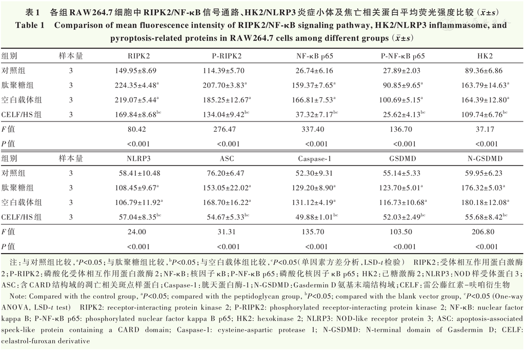
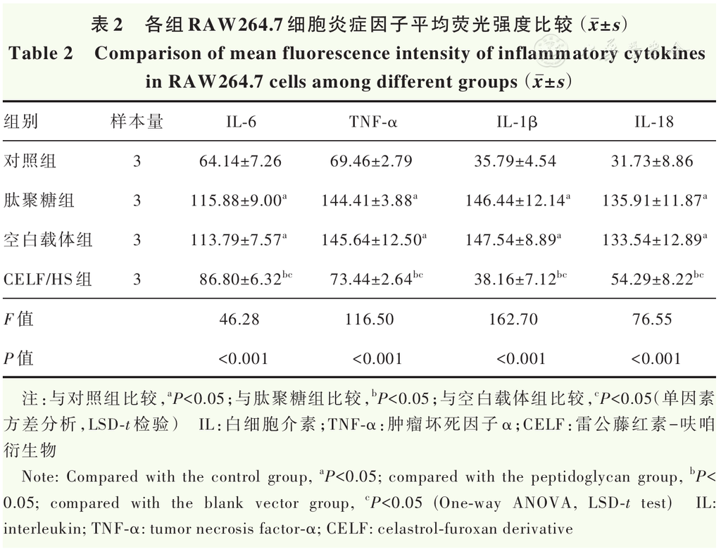
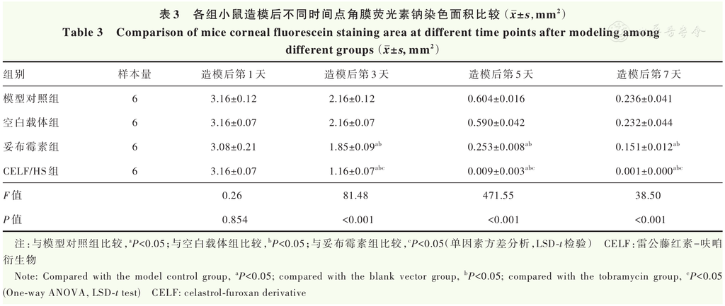
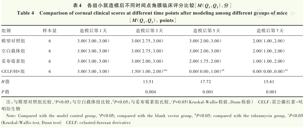
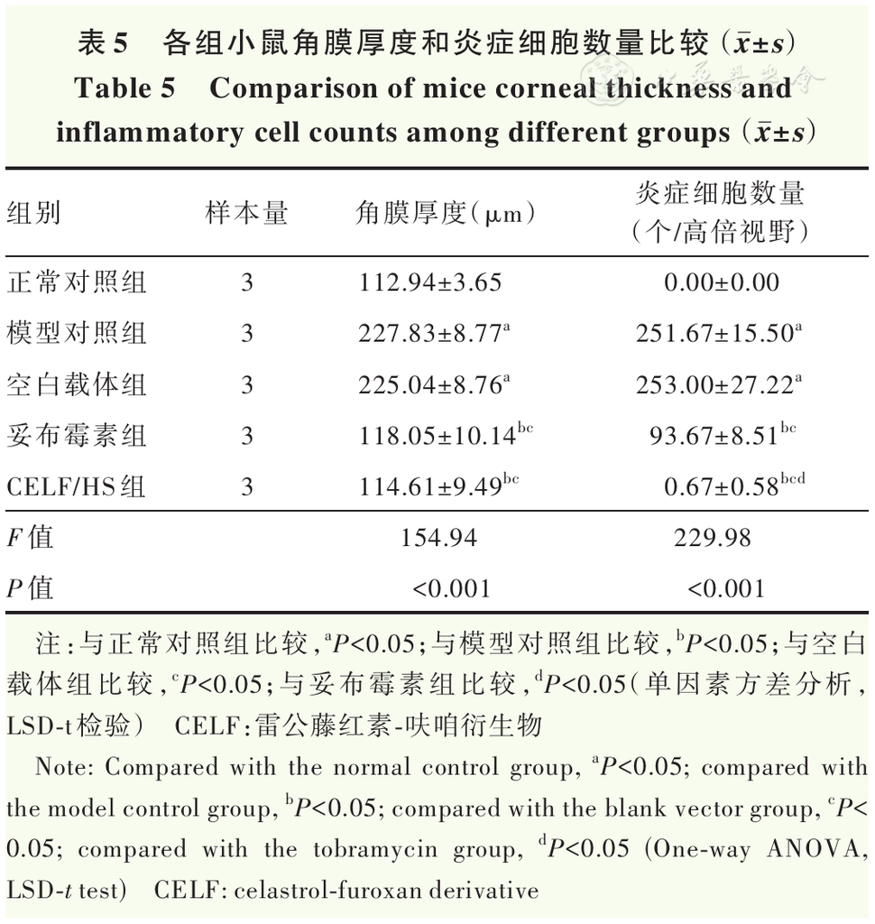
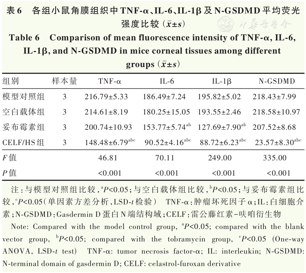
Results The in vitro plate assay showed that CELF/HS at 6.25 μg/ml effectively inhibited bacterial growth after 8 hours of incubation. Scanning electron microscopy and live/dead bacterial staining further demonstrated that CELF/HS at 6.25 μg/ml effectively killed SA. Compared with the PGN group, the CELF/HS group showed significantly lower fluorescence intensities of RIPK2/NF-κB signaling pathway-related proteins (P-RIPK2, RIPK2, P-NF-κB p65, and NF-κB p65), HK2/NLRP3 signaling axis-related proteins (HK2, NLRP3, ASC, and caspase-1), pyroptosis-related proteins (GSDMD and N-GSDMD), and inflammatory cytokines (IL-6, TNF-α, IL-1β, and IL-18) in RAW264.7 cells, with statistically significant differences (all P<0.05). With increasing observation time, corneal opacity, edema, and fluorescein sodium-stained area gradually decreased in the CELF/HS group, with marked improvement in corneal transparency on day 7. On days 3, 5, and 7 after model establishment, the corneal fluorescein sodium-stained area in the CELF/HS group was significantly smaller than those in the model control, blank vector, and tobramycin groups (all P<0.05). On day 7, the colony counts of re-cultured infected corneal tissues were (11 170±245), (11 047±925), (6 567±404), and (58.33±15.28) CFU in the model control, blank vector, tobramycin, and CELF/HS groups, respectively, with a significant overall difference (F=297.60, P<0.001). Colony counts were significantly lower in the tobramycin group than in the model control and blank vector groups, and were significantly lower in the CELF/HS group than in the model control, blank vector, and tobramycin groups (all P<0.05). Corneal thickness was significantly greater in the model control and blank vector groups than in the normal control, tobramycin, and CELF/HS groups (all P<0.05). The number of inflammatory cells was significantly lower in the tobramycin group than in the model control and blank vector groups, and was significantly lower in the CELF/HS group than in the model control, blank vector, and tobramycin groups (all P<0.05). The mean fluorescence intensities of TNF-α, IL-6, IL-1β, and N-GSDMD in corneal tissues were significantly lower in the CELF/HS group than in the model control, blank vector, and tobramycin groups (all P<0.05).
Conclusions CELF/HS effectively kills SA while alleviating corneal inflammation. Its therapeutic effects may be associated with the inhibition of the HK2/NLRP3 and RIPK2/NF-κB signaling pathways, reduction of NLRP3 inflammasome formation, and suppression of GSDMD-mediated pyroptosis.
KEYWORDS:
Celastrol;Furoxan derivative;Bacterial keratitis;Pyroptosis;Inflammasome
COPYRIGHTS:
Copyright by Chinese Medical Association
No content published by the journals of Chinese Medical Association may be reproduced or abridged without authorization. Please do not use or copy the layout and design of the journals without permission.
All articles published represent the opinions of the authors, and do not reflect the official policy of the Chinese Medical Association or the Editorial Board, unless this is clearly specified.
Authors Info & Affiliations
Wang Mengke
People’s Hospital of Zhengzhou University, Department of Ophthalmology, Henan Provincial People’s Hospital, Henan Eye Hospital, Henan Key Laboratory of Ophthalmology and Visual Science, Zhengzhou 450003, China
Liu Kang
People’s Hospital of Zhengzhou University, Department of Ophthalmology, Henan Provincial People’s Hospital, Henan Eye Hospital, Henan Key Laboratory of Ophthalmology and Visual Science, Zhengzhou 450003, China
Wang Wang
People’s Hospital of Zhengzhou University, Department of Ophthalmology, Henan Provincial People’s Hospital, Henan Eye Hospital, Henan Key Laboratory of Ophthalmology and Visual Science, Zhengzhou 450003, China
Feng Huayang
People’s Hospital of Zhengzhou University, Department of Ophthalmology, Henan Provincial People’s Hospital, Henan Eye Hospital, Henan Key Laboratory of Ophthalmology and Visual Science, Zhengzhou 450003, China
Li Jiake
People’s Hospital of Zhengzhou University, Department of Ophthalmology, Henan Provincial People’s Hospital, Henan Eye Hospital, Henan Key Laboratory of Ophthalmology and Visual Science, Zhengzhou 450003, China
Deng Yuting
People’s Hospital of Zhengzhou University, Department of Ophthalmology, Henan Provincial People’s Hospital, Henan Eye Hospital, Henan Key Laboratory of Ophthalmology and Visual Science, Zhengzhou 450003, China
Li Jingguo
People’s Hospital of Zhengzhou University, Department of Ophthalmology, Henan Provincial People’s Hospital, Henan Eye Hospital, Henan Key Laboratory of Ophthalmology and Visual Science, Zhengzhou 450003, China
Li Zhanrong
People’s Hospital of Zhengzhou University, Department of Ophthalmology, Henan Provincial People’s Hospital, Henan Eye Hospital, Henan Key Laboratory of Ophthalmology and Visual Science, Zhengzhou 450003, China
Figures & Tables
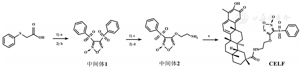
Figure 1 Synthesis of CELF a: 30% H2O2, glacial acetic acid, room temperature, 3 hours; b: fuming HNO3, glacial acetic acid, 100°C, 4 hours; c: N-Boc-ethanolamine, NaOH (aq), THF, room temperature, 1 hour; d: HCl/EtOAc, DCM, room temperature, 3 hours; e: EDCI, HOBt, DIPEA, DCM, room temperature, 1 hour CELF: celastrol-furoxan derivative; THF: tetrahydrofuran; EtOAc: ethyl acetate; DCM: dichloromethane; EDCI: 1-ethyl-3-(3-dimethylaminopropyl) carbodiimide hydrochloride; HOBt: 1-hydroxybenzotriazole; DIPEA: N,N-diisopropylethylamine
▼click to see the rest
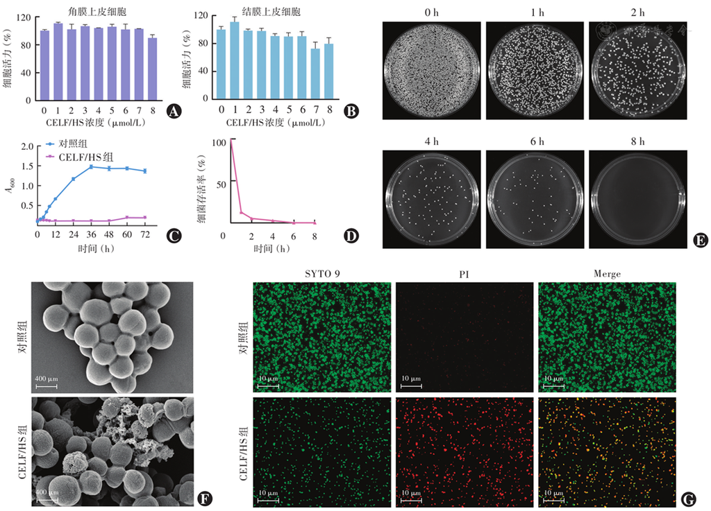
Figure 2 Biological safety and antibacterial activity of CELF/HS in vitro A, B: The biological safety of different concentrations of CELF/HS on corneal epithelial cells and conjunctival epithelial cells C: Crowth curve of SA with different treatments D: SA survival ratio changes at different time points of co-culture E: The antibacterial effect of CELF/HS treated at different time points by bacterial plate counting assay F: Electron microscopy images of SA with different treatments (scale bar=400 nm) G: Fluorescent live/dead images of SA with different treatments (scale bar=10 μm) CELF: celastrol-furoxan derivatives; SA: Staphylococcus aureus
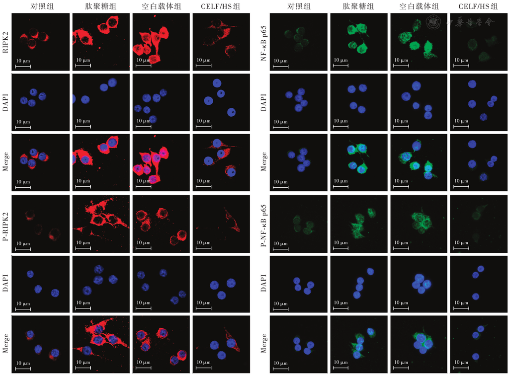
Figure 3 Representative immunofluorescence staining images of RIPK2, P-RIPK2, NF-κB p65, and P-NF-κB p65 in RAW264.7 cells from different groups (scale bar=10 μm) RIPK2: receptor-interacting protein kinase 2; P-RIPK2: phosphorylated receptor-interacting protein kinase 2; NF-κB: nuclear factor kappa B; P-NF-κB p65: phosphorylated nuclear factor kappa B p65; CELF: celastrol-furoxan derivative
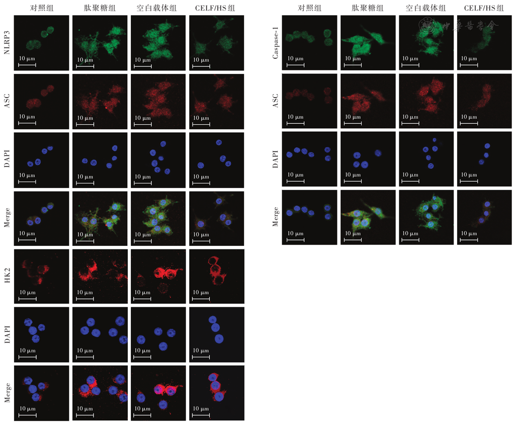
Figure 4 Representative immunofluorescence staining images of NLRP3, ASC, caspase-1, and HK2 in RAW264.7 cells from different groups (scale bar=10 μm) NLRP3: NOD-like receptor protein 3; ASC: apoptosis-associated speck-like protein containing a CARD domain; Caspase-1: cysteine-aspartic protease 1; HK2: hexokinase 2; CELF: celastrol-furoxan derivative
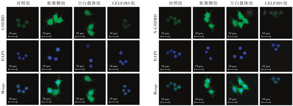
Figure 5 Representative immunofluorescence staining images of GSDMD and N-GSDMD in RAW264.7 cells from different groups (scale bar=10 μm) N-GSDMD: N-terminal domain of Gasdermin D; CELF: celastrol-furoxan derivative
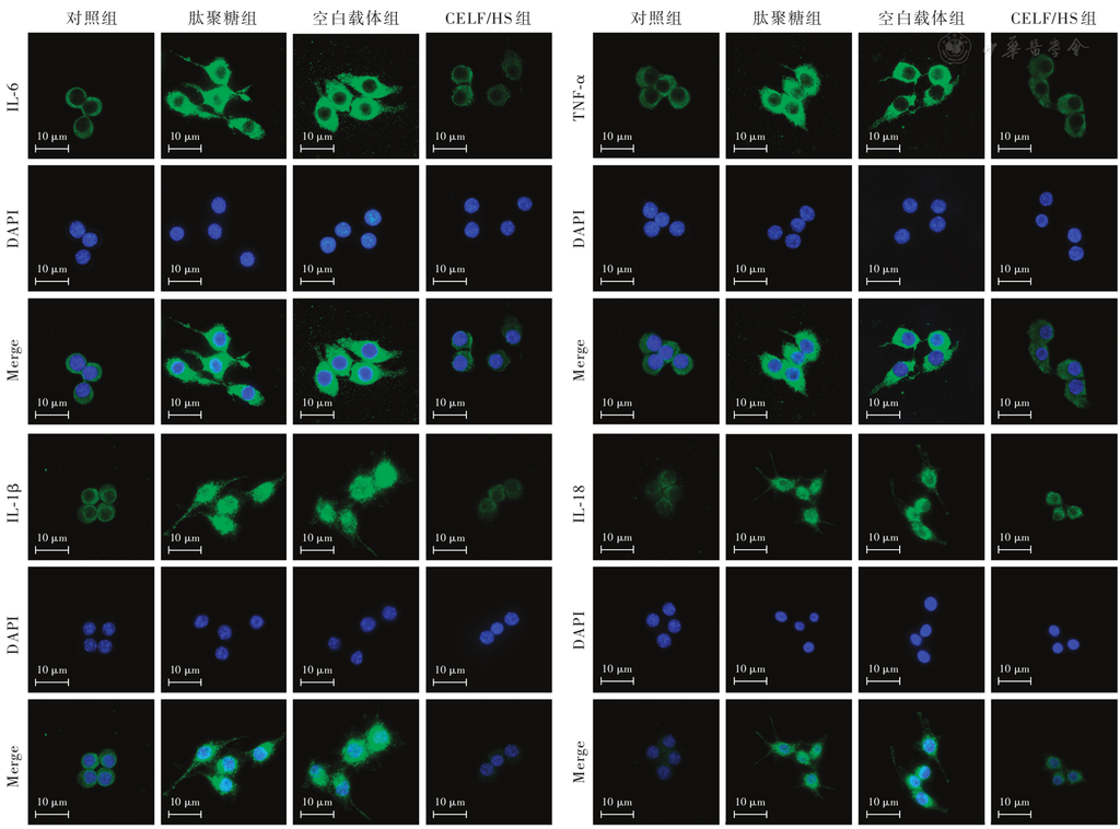
Figure 6 Representative immunofluorescence staining images of IL-6, TNF-α, IL-1β, and IL-18 in RAW264.7 cells from different groups (scale bar=10 μm) IL: interleukin; TNF-α: tumor necrosis factor-α; CELF: celastrol-furoxan derivative
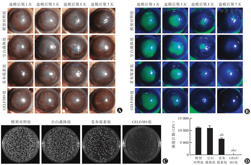
Figure 7 Therapeutic effect of CELF/HS on bacterial keratitis in vivo A: Diffuse light (×25) under slit lamp microscope B: Fluorescein sodium stained images under slit lamp microscope (×25) C: Photos of TSA plates for re-culturing the infected corneal tissues on the 7th day of different treatments D: Comparison of colony counts from re-cultured tissues among different groups F=297.60,P<0.001; Compared with the model control group, aP<0.05; compared with the blank vector group, bP<0.05; compared with the tobramycin group, cP<0.05 (One-way ANOVA, Tukey test) CELF: celastrol-furoxan derivative; TSA: tryptic soy agar; CFU: colony-forming unit
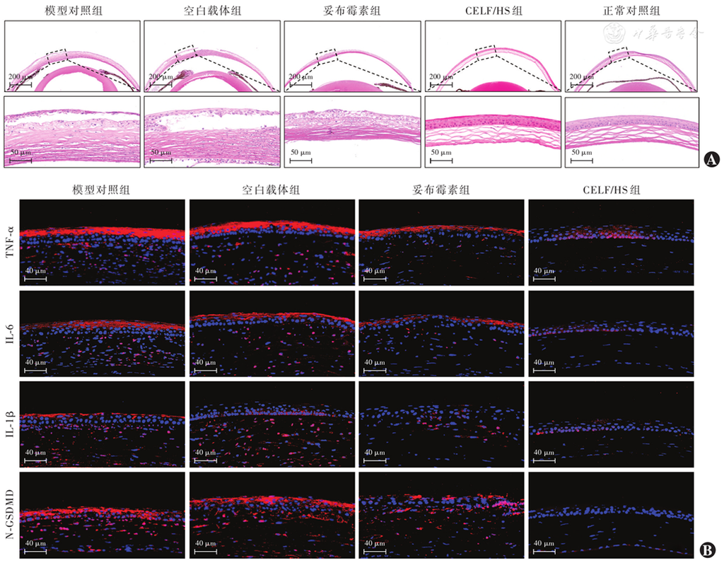
Figure 8 Histological evaluation of mice corneal tissues on day 7 after modeling A: Representative hematoxylin-eosin staining images (scale bar=200 μm) The lower figures showed local magnification of dashed box (scale bar=50 μm) B: Representative immunofluorescence images of TNF-α, IL-6, IL-1β, and N-GSDMD (scale bar=40 μm) Nuclei were counterstained with DAPI (blue), and the target proteins were shown in red TNF-α: tumor necrosis factor-α; IL: interleukin; N-GSDMD: N-terminal domain of gasdermin D; CELF: celastrol-furoxan derivative
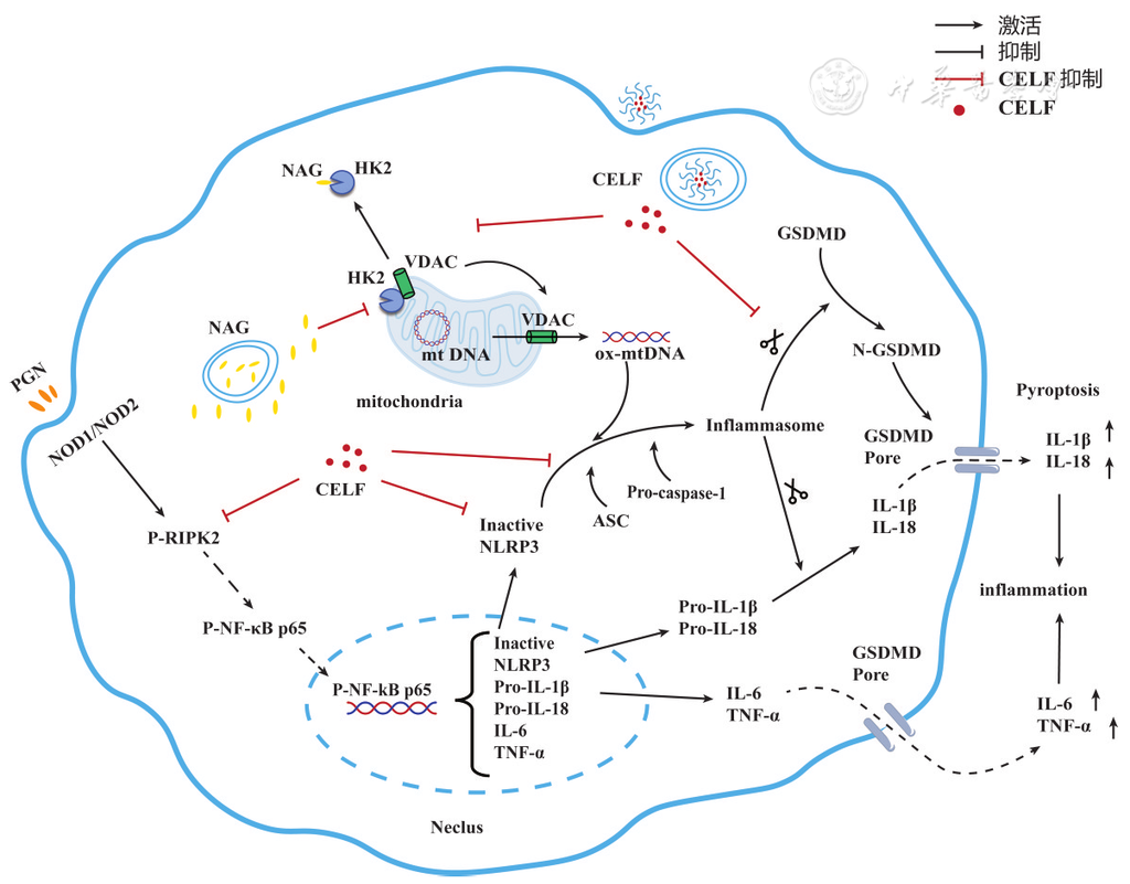
Figure 9 Proposed mechanism underlying the therapeutic effects of CELF/HS against Staphylococcus aureus-induced bacterial keratitis P-RIPK2: phosphorylated receptor-interacting protein kinase 2; P-NF-κB p65: phosphorylated nuclear factor κB p65; HK2: hexokinase 2; NLRP3: NOD-like receptor protein 3; ASC: apoptosis-associated speck-like protein containing a CARD domain; Caspase-1: cysteine-aspartic protease 1; N-GSDMD: N-terminal domain of Gasdermin D; IL: interleukin; TNF-α: tumor necrosis factor-α; CELF: celastrol-furoxan derivative
References
[1] Lin A, Rhee MK, Akpek EK, et al. Bacterial keratitis preferred practice pattern®[J]. Ophthalmology, 2019, 126(1):P1-P55. DOI: 10.1016/j.ophtha.2018.10.018.
▼click to see more references
[2] Shah S, Wozniak R. Staphylococcus aureus and Pseudomonas aeruginosa infectious keratitis: key bacterial mechanisms that mediate pathogenesis and emerging therapeutics[J]. Front Cell Infect Microbiol, 2023, 13:1250257. DOI: 10.3389/fcimb.2023.1250257.
[3] Yan C, Zhao H, Pan Q, et al. Novel treatments for bacterial keratitis: a review[J]. Int J Pharm, 2025, 681:125793. DOI: 10.1016/j.ijpharm.2025.125793.
[4] Tuft S, Somerville TF, Li JO, et al. Bacterial keratitis: identifying the areas of clinical uncertainty[J]. Prog Retin Eye Res, 2022, 89:101031. DOI: 10.1016/j.preteyeres.2021.101031.
[5] Li Y, Gao W, Qiu Y, et al. RING1 dictates GSDMD-mediated inflammatory response and host susceptibility to pathogen infection[J]. Cell Death Differ, 2025, 32(11):2066-2077. DOI: 10.1038/s41418-025-01527-2.[6] Chai Q, Yu S, Zhong Y, et al. A bacterial phospholipid phosphatase inhibits host pyroptosis by hijacking ubiquitin[J]. Science, 2022, 378(6616):eabq0132. DOI: 10.1126/science.abq0132.
[7] Barnett KC, Li S, Liang K, et al. A 360° view of the inflammasome: mechanisms of activation, cell death, and diseases[J]. Cell, 2023, 186(11):2288-2312. DOI: 10.1016/j.cell.2023.04.025.
[8] Larochelle J, Tishko RJ, Yang C, et al. Receptor-interacting protein kinase 2 (RIPK2) profoundly contributes to post-stroke neuroinflammation and behavioral deficits with microglia as unique perpetrators[J]. J Neuroinflammation, 2023, 20(1):221. DOI: 10.1186/s12974-023-02907-6.
[9] Jing G, Gong H, Wang H, et al. OTUD1 exacerbates sepsis-associated encephalopathy by promoting HK2 mitochondrial release to drive microglia pyroptosis[J]. J Neuroinflammation, 2025, 22(1):154. DOI: 10.1186/s12974-025-03480-w.
[10] Baik SH, Ramanujan VK, Becker C, et al. Hexokinase dissociation from mitochondria promotes oligomerization of VDAC that facilitates NLRP3 inflammasome assembly and activation[J]. Sci Immunol, 2023, 8(84):eade7652. DOI: 10.1126/sciimmunol.ade7652.
[11] Xu S, Feng Y, He W, et al. Celastrol in metabolic diseases: progress and application prospects[J]. Pharmacol Res, 2021, 167:105572. DOI: 10.1016/j.phrs.2021.105572.
[12] Meng Z, Liu Q, Chen H, et al. Therapeutic potential of celastrol in bacterial infections: current research advancements and future perspectives[J]. Pharmacol Res, 2025, 216:107774. DOI: 10.1016/j.phrs.2025.107774.
[13] Jing M, Yang J, Zhang L, et al. Celastrol inhibits rheumatoid arthritis through the ROS-NF-κB-NLRP3 inflammasome axis[J]. Int Immunopharmacol, 2021, 98:107879. DOI: 10.1016/j.intimp.2021.107879.
[14] Li SM, Chou JY, Tsai SE, et al. Synthesis and anti-inflammatory activity evaluation of NO-releasing furoxan/1,2,4-triazole hybrid derivatives[J]. Eur J Med Chem, 2023, 257:115496. DOI: 10.1016/j.ejmech.2023.115496.
[15] Geng W, Liu X, Tao B, et al. Nitric oxide scavenging and hydrogen sulfide production synergistically treat rheumatoid arthritis[J]. Adv Healthc Mater, 2023, 12(4):e2202380. DOI: 10.1002/adhm.202202380.
[16] Ning Q, Sun X, Cui H, et al. Dual-corn-derived nanofiber membrane for subconjunctival injury: sequential release of dual-natural products for programmed anti-inflammation and anti-fibrosis[J]. J Control Release, 2025, 381:113577. DOI: 10.1016/j.jconrel.2025.02.073.
[17] Dai Y, Zhang J, Zhang S, et al. Ag/Cu nanoparticles-loaded glycocalyx biomimetic corneal bandage lenses for combatting bacterial keratitis[J]. J Control Release, 2024, 376:382-394. DOI: 10.1016/j.jconrel.2024.10.015.
[18] Carion TW, Kracht D, Strand E, et al. VIP modulates the ALX/FPR2 receptor axis toward inflammation resolution in a mouse model of bacterial keratitis[J]. Prostaglandins & Other Lipid Mediators, 2019, 140:18-25. DOI: 10.1016/j.prostaglandins.2018.12.001.
[19] Man MQ, Wakefield JS, Mauro TM, et al. Regulatory role of nitric oxide in cutaneous inflammation[J]. Inflammation, 2022, 45(3):949-964. DOI: 10.1007/s10753-021-01615-8.
[20] Liu X, Zhang Z, Ruan J, et al. Inflammasome-activated gasdermin D causes pyroptosis by forming membrane pores[J]. Nature, 2016, 535(7610):153-158. DOI: 10.1038/nature18629.
[21] Chen H, Zhang J, He Y, et al. Exploring the role of Staphylococcus aureus in inflammatory diseases[J]. Toxins (Basel), 2022, 14(7):464. DOI: 10.3390/toxins14070464.
[22] Vande Walle L, Lamkanfi M. Drugging the NLRP3 inflammasome: from signalling mechanisms to therapeutic targets[J]. Nat Rev Drug Discov, 2024, 23(1):43-66. DOI: 10.1038/s41573-023-00822-2.
[23] Maran JJ, Adesina MM, Green CR, et al. The central role of the NLRP3 inflammasome pathway in the pathogenesis of age-related diseases in the eye and the brain[J]. Ageing Res Rev, 2023, 88:101954. DOI: 10.1016/j.arr.2023.101954.
[24] Xu Y, Lin F, Liao G, et al. Ripks and neuroinflammation[J]. Mol Neurobiol, 2024, 61(9):6771-6787. DOI: 10.1007/s12035-02403981-4.
[25] Cheng X, Barakat R, Pavani G, et al. Nod1-dependent NF-kB activation initiates hematopoietic stem cell specification in response to small Rho GTPases[J]. Nat Commun, 2023, 14(1):7668. DOI: 10.1038/s41467-023-43349-1.