Authors:Fan Yuanyuan,Luo Yufan, Xie Ping, Hu Zizhong
Corresponding author: Hu Zizhong, Email: huzizhong@njmu.edu.cn
Published:2026-09-10
DOI:10.3760/cma.j.cn115989-20260427-00191
ABSTRACT
Objective To construct engineered exosomes loaded with the p300 inhibitor A485 (A485@EV) and to investigate their effects and mechanisms in inhibiting hypoxia-induced pathological neovascularization by targeting inhibition of lactylation.
Methods A485@EV was prepared by sonication, and its markers, morphology and physical and chemical properties were identified. The encapsulation efficiency, loading efficiency and release profile of A485 were determined. Human umbilical vein endothelial cells (HUVECs) and human microglial cell line 3 (HMC3) were divided into control group, free A485 group, and A485@EV group, and cultured under 1% O 2 for 24 hours to establish a hypoxia model. Then equal volumes of phosphate buffered saline (PBS), free A485 or A485@EV were added to the cell culture system correspondingly, so that the final concentration of free A485 and A485@EV was 20 μmol/L and 20 μg/ml, respectively. EdU proliferation assay, Transwell migration assay, and tube formation assay were used to evaluate the proliferation, migration, and tube formation of HUVECs. EdU proliferation assay, Transwell migration assay, wound healing assay, and Western blot were used to evaluate the proliferation, migration, and wound healing of HMC3 as well as the cyclooxygenase-2 (COX-2) protein expression. Eighteen healthy 7-day-old male C57BL/6J mice were selected to establish an oxygen-induced retinopathy (OIR) model. The OIR mice were randomly divided into control group, free A485 group, and A485@EV group using the random number table method, with 6 mice in each group and mice received an intravitreal injection of 1 μl of PBS, 1 mmol A485, or 1 μg A485@EV, respectively. Avascular area, neovascular area, and the lactylation levels of retinal endothelial cells and microglia were detected by immunofluorescent imaging. The study protocol was approved by the Animal Ethics Committee of Nanjing Medical University (No. IACUC-2202032-1).
Results The successfully constructed A485@EV exhibited a typical cup-shaped morphology, with a mean particle size of (167.0±70.6)nm, a zeta potential of -35.63 mV, an encapsulation efficiency of (40.91±2.30)%, a loading efficiency of (20.45±1.15)%, and a 24-hour cumulative release rate of approximately 73.1%. Compared with the control group and free A485 group, the EdU positive rate, the number of migrating cells and the tube formation ability of HUVECs in A485@EV group were significantly reduced (all P<0.05). Compared with the control group and free A485 group, the EdU positive rate, the number of migrating cells, scratch healing rate and relative expression of COX-2 protein in HMC3 cells were also significantly reduced (all P<0.05). In the OIR mouse model, avascular and neovascular areas were both lower in the A485@EV group than in the control and free A485 groups, with statistically significant differences (all P<0.05). IB4-positive endothelial cells and IBA1-positive microglia showed markedly weaker fluorescence of lactylation in the A485@EV group than in the control and free A485 groups.
Conclusions Engineered exosome loaded with A485 may attenuate hypoxia-induced endothelial cell tube formation and microglial activation by targeting inhibition of the lactylation effect, thereby inhibiting pathological neovascularization.
KEYWORDS:
Pathological neovascularization;Engineered exosomes;A485;Lactylation
COPYRIGHTS:
Copyright by Chinese Medical Association
No content published by the journals of Chinese Medical Association may be reproduced or abridged without authorization. Please do not use or copy the layout and design of the journals without permission.
All articles published represent the opinions of the authors, and do not reflect the official policy of the Chinese Medical Association or the Editorial Board, unless this is clearly specified.
Authors Info & Affiliations
Fan Yuanyuan
Department of Ophthalmology, The First Affiliated Hospital of Nanjing Medical University, Nanjing 210029, China
Luo Yufan
Department of Ophthalmology, The First Affiliated Hospital of Nanjing Medical University, Nanjing 210029, China
Xie Ping
Department of Ophthalmology, The First Affiliated Hospital of Nanjing Medical University, Nanjing 210029, China
Hu Zizhong
Department of Ophthalmology, The First Affiliated Hospital of Nanjing Medical University, Nanjing 210029, China
Figures & Tables
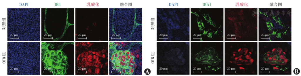
Figure 1 Lactylation levels in mice retinal endothelial cells and microglia in each group (Scale bar=20 μm) A: Co-localization of endothelial cells (IB4, green) and pan-lactylation (red) B: Co-localization of microglia (IBA1, green) and pan-lactylation (red) OIR: oxygen-induced retinopathy
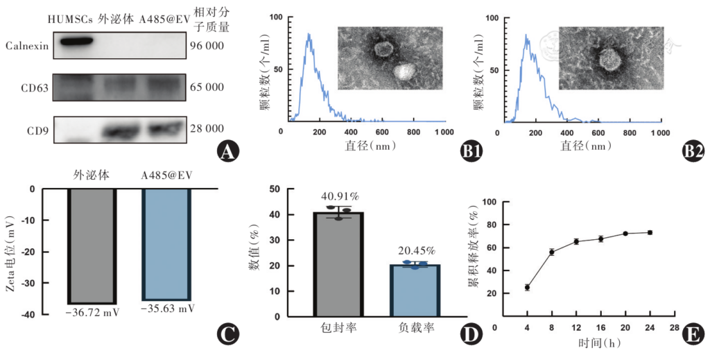
Figure 2 Identification of exosomes and construction and characterization of engineered exosomes (A485@EV) A: Electrophoretogram of markers expression of exosomes and A485@EV CD63 and CD9 proteins were highly expressed and the endoplasmic reticulum marker Calnexin was not obviously expressed B: Size distribution and TEM images of exosomes and A485@EV (Scale bar=100 nm) B1: exosomes B2: A485@EV C: Average zeta potential of exosomes and A485@EV D: Encapsulation efficiency and loading efficiency of A485@EV E: Release profile of A485@EV HUMSCs: human umbilical cord mesenchymal stem cells; TEM: transmission electron microscopy
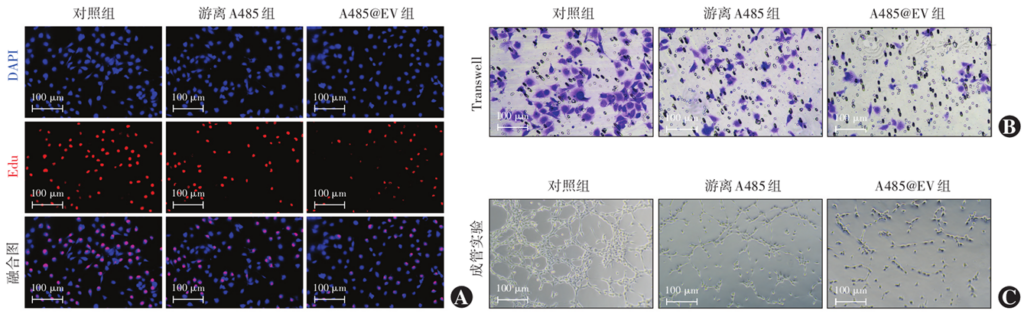
Figure 3 Proliferation, migration, and tube formation of different HUVECs group A: Staining images of EdU proliferation assay (Scale bar=100 µm) B: Representative images of Transwell migration assay (crystal violet, Scale bar=100 µm) C: Representative images of tube formation assay (Scale bar=100 µm) HUVECs: human umbilical vein endothelial cells
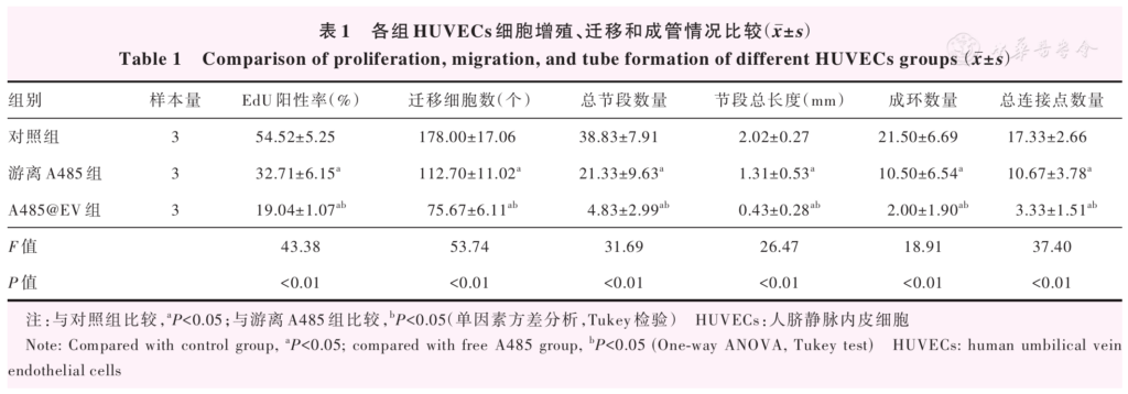
Table 1 Comparison of proliferation, migration, and tube formation of different HUVECs groups (mean±SD) Note: Compared with control group, a P<0.05; compared with free A485 group, b P<0.05 (One-way ANOVA, Tukey test) HUVECs: human umbilical vein endothelial cells
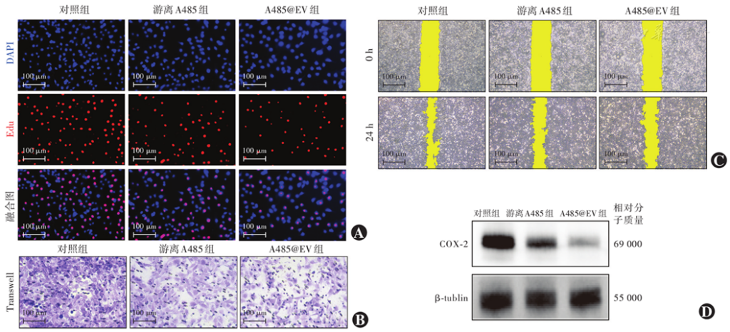
Figure 4 Proliferation, migration, wound healing, and COX-2 expression in different HMC3 groups A: Staining images of EdU proliferation assay (Scale bar=100 µm) B: Representative images of Transwell migration assay (crystal violet, Scale bar=100 µm) C: Representative images of wound healing assay (Scale bar=100 µm) D: Electrophoretogram of COX-2 protein expression HMC3: human microglial cell line 3; COX-2: cyclooxygenase-2
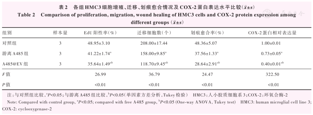
Table 2 Comparison of proliferation, migration, wound healing of HMC3 cells and COX-2 protein expression among different groups (mean±SD) Note: Compared with control group, a P<0.05; compared with free A485 group, b P<0.05 (One-way ANOVA, Tukey test) HMC3: human microglial cell line 3; COX-2: cyclooxygenase-2
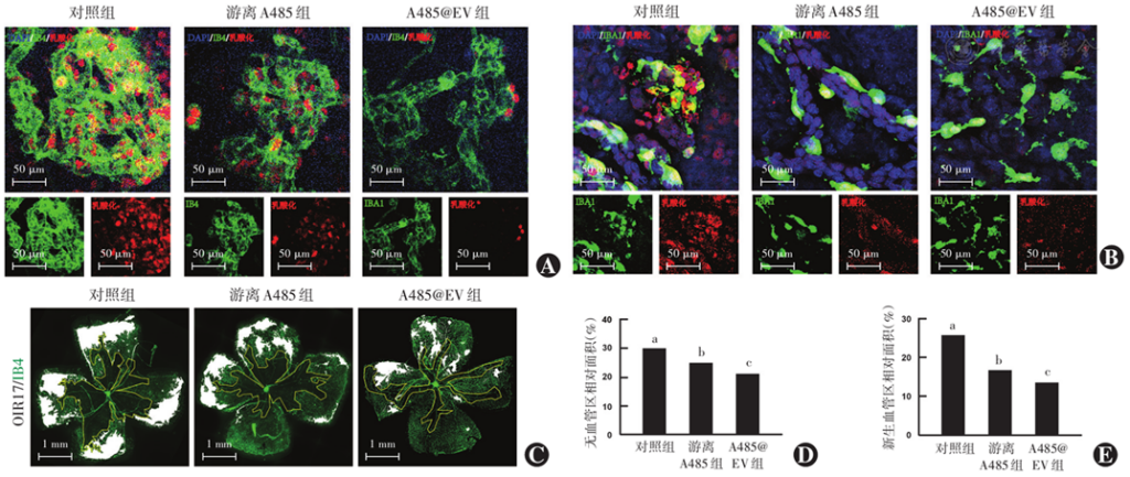
Figure 5 Comparison of the degree of lactylation and the areas of non-vascular and neovascular regions in OIR mice retina among different groups A: Co-localization of endothelial cells (IB4, green) and pan-lactylation (red) (Scale bar=50 µm) B: Co-localization of microglia (IBA1, green) and pan-lactylation (red) (Scale bar=50 µm) C: Avascular areas (outlined in yellow lines) and neovascular areas (covered in white shadow) (Scale bar=1 mm) D: Comparison of retinal avascular area F=21.98, P<0.05. Compared with control group, aP<0.05; compared with free A485 group, bP<0.05 (One-way ANOVA, Tukey test; n=6) E: Comparison of retinal neovascular area F=59.59, P<0.05. Compared with control group, aP<0.05; compared with free A485 group, bP<0.05 (One-way ANOVA, Tukey test; n=6) OIR: oxygen-induced retinopathy
References click to collapse
[1] Sheth JU, Stewart MW, Narayanan R, et al. Macular neovascularization[J]. Surv Ophthalmol, 2025, 70(4):653-675. DOI: 10.1016/j.survophthal.2024.08.003.
[2] Ng D, Ruamviboonsuk P, Apte RS, et al. International consensuses and controversies on causes, diagnosis and management of diabetic macular edema(DME)[J].ProgRetin EyeRes,2025,109:101406.DOI: 10.1016/j.preteyeres.2025.101406.
[3] Xu M, Fan R, Fan X, et al. Progress and challenges of anti-VEGF agents and their sustained-release strategies for retinal angiogenesis[J]. Drug Des Devel Ther, 2022, 16:3241-3262. DOI: 10.2147/DDDT.S383101.
[4] Chen DY, Sun NH, Chen X, et al. Endothelium-derived semaphorin 3G attenuates ischemic retinopathy by coordinating β-catenin-dependent vascular remodeling[J/OL]. J Clin Invest, 2021, 131(4):e135296[2026-04-22]. http://www.ncbi.nlm.nih.gov/pubmed/33586674. DOI: 10.1172/JCI135296.
[5] Zhao B, Zhao Y, Sun X. Mechanism and therapeutic targets of circulating immune cells in diabetic retinopathy[J]. Pharmacol Res, 2024, 210:107505. DOI: 10.1016/j.phrs.2024.107505.
[6] Hu A, Schmidt M, Heinig N. Microglia in retinal angiogenesis and diabetic retinopathy[J]. Angiogenesis, 2024, 27(3):311-331. DOI: 10.1007/s10456-024-09911-1.
[7] Chai P, Zhao F, Jia R, et al. Lactate/lactylation in ocular development and diseases[J]. Trends Mol Med, 2025, 31(12):1073-1076. DOI: 10.1016/j.molmed.2024.07.001.
[8] 高艳, 茅希颖,陈明康,等.乳酸在视网膜病变中的作用及机制研究进展[J]. 中华眼底病杂志,2024, 40(12):975-979. DOI: 10.3760/cma.j.cn511434-20240731-00292. Gao Y, Mao XY, Chen MK, et al. Research progress on the role and mechanism of lactate in retinal diseases[J]. Chin J Ocul Fundus Dis, 2024, 40(12):975-979. DOI: 10.3760/cma.j.cn511434-20240731-00292.
[9] Wang X, Fan W, Li N, et al. YY1 lactylation in microglia promotes angiogenesis through transcription activation-mediated upregulation of FGF2[J]. Genome Biol, 2023, 24(1):87. DOI: 10.1186/s13059-023-02931-y.
[10] Huang J, Wang X, Li N, et al. YY1 Lactylation aggravates autoimmune uveitis by enhancing microglial functions via inflammatory genes[J/OL]. Adv Sci (Weinh), 2024, 11(19):e2308031[2026-04-22]. http://www.ncbi.nlm.nih.gov/pubmed/38493498. DOI: 10.1002/advs.202308031.
[11] Fan W, Zeng S, Wang X, et al. A feedback loop driven by H3K9 lactylation and HDAC2 in endothelial cells regulates VEGF-induced angiogenesis[J]. Genome Biol, 2024, 25(1):165. DOI: 10.1186/s13059-024-03308-5.
[12] Wang X, Huang J, Fan W, et al. Lactate-binding protein DNMT3A in HRMECs promotes angiogenesis by upregulating VEGFA through HIF-1α lactylation[J]. Genome Biol, 2025, 26(1):377. DOI: 10.1186/s13059-025-03845-7.
[13] Zuo S, Li L, Tang J, et al. Lactylation-boosted m5C RNA modification drives choroidal neovascularization[J]. Research (Wash D C), 2025, 8: 0913. DOI: 10.34133/research.0913.
[14] Zhang D, Tang Z, Huang H, et al. Metabolic regulation of gene expression by histone lactylation[J]. Nature, 2019, 574(7779):575-580. DOI: 10.1038/s41586-019-1678-1.
[15] 王梦华, 胡芷柔.外泌体共载牛血清白蛋白联合异槲皮苷纳米体系对高糖诱导人RPE细胞的保护作用及机制[J].中华实验眼科杂志, 2025,43(9):794-800.DOI: 10.3760/cma.j.cn115989-20250515-00157. Wang MH, Hu ZR. Protective effects and mechanisms of exosomes co-loaded with BSA@ISO nanoparticles on high glucose-induced human retinal pigment epithelial cells[J]. Chin J Exp Ophthalmol, 2025, 43(9):794-800. DOI: 10.3760/cma.j.cn115989-20250515-00157.
[16] Fan Y, Ge P, Wang X, et al. Microglia-specific interleukin-4 delivery by engineered extracellular vesicles restores inner blood-retinal barrier in diabetic retinopathy via GAS6-MERTK pathway[J]. J Nanobiotechnology, 2025, 24(1):101. DOI: 10.1186/s12951-025-03976-w.
[17] Hu Z, Wang J, Pan T, et al. The exosome-transmitted lncRNA LOC100132249 induces endothelial dysfunction in diabetic retinopathy[J]. Diabetes, 2023, 72(9):1307-1319. DOI: 10.2337/db22-0435.
[18] Ge P, Wang J, Xie P, et al. Exosomes in treating eye diseases: targeting strategies[J]. Life Sci, 2025, 371:123598. DOI: 10.1016/j.lfs.2025.123598.
[19] Massoumi H, Katz EA, Alviar M, et al. Corneal nerve regeneration via MSC-derived EVs: tissue source and culture dimensionality dictate miRNA cargo and therapeutic efficacy[J/OL]. Small, 2026, 22(6): e07451[2026-04-22]. http://www.ncbi.nlm.nih.gov/pubmed/41340474. DOI: 10.1002/smll.202507451.
[20] Zhou H, Liu Y, Zhou T, et al. IL-23 priming enhances the neuroprotective effects of MSC-derived exosomes in treating retinal degeneration[J]. Invest Ophthalmol Vis Sci, 2024, 65(10):8. DOI: 10.1167/iovs.65.10.8.
[21] Fan R, Su L, Zhang H, et al. Enhanced therapeutic effect of PEDF-loaded mesenchymal stem cell-derived small extracellular vesicles against oxygen-induced retinopathy through increased stability and penetrability of PEDF[J]. J Nanobiotechnology, 2023, 21(1):327. DOI: 10.1186/s12951-023-02066-z.
[22] Clements JN, Mesawich J, Twisdale C. Current and emerging pharmacologic therapies for diabetic retinopathy[J/OL]. Pharmacotherapy, 2026, 46(3):e70119[2026-04-24]. http://www.ncbi.nlm.nih.gov/pubmed/41693076. DOI: 10.1002/phar.70119.
[23] Guan J, Meng F, Wang C, et al. Recent advances in engineered exosome-based therapies for ocular vascular disease[J]. J Nanobiotechnology, 2025, 23(1):526. DOI: 10.1186/s12951-025-03589-3.
[24] Wu Y, Wang J, Pan T, et al. Human lens epithelial-secreted exosomes attenuate ocular angiogenesis via inhibiting microglial activation[J]. Exp Eye Res, 2024, 241:109837. DOI: 10.1016/j.exer.2024.109837.
[25] 牛瑞姣, 刘锦荟, 董怡,等.选择性脑亚低温对脑缺血再灌注损伤大鼠p300及乳酸化修饰的影响[J].中华危重病急救医学,2026,38(2): 152-157. DOI: 10.3760/cma.j.cn121430-20240824-00414. Niu RJ, Liu JH, Dong Y, et al. Effect of selective cerebral mild hypothermia on p300 and lactylation modification in rats following cerebral ischemic reperfusion injury[J]. Chin Crit Care Med, 2026, 38(2):152-157. DOI: 10.3760/cma.j.cn121430-20240824-00414.
[26] Lan W, Chen X, Yu H, et al. UGDH lactylation aggravates osteoarthritis by suppressing glycosaminoglycan synthesis and orchestrating nucleocytoplasmic transport to activate MAPK signaling[J/OL]. Adv Sci (Weinh), 2025, 12(20):e2413709[2026-04-24]. http://www.ncbi.nlm.nih.gov/pubmed/40150862. DOI: 10.1002/advs.202413709.
[27] Deng J, Li Y, Yin L, et al. Histone lactylation enhances GCLC expression and thus promotes chemoresistance of colorectal cancer stem cells through inhibiting ferroptosis[J]. Cell Death Dis, 2025, 16(1): 193. DOI: 10.1038/s41419-025-07498-z.