Authors:Ren Yaoyao, Liu Yang
Corresponding author: Liu Yang, Email:drliuyang_eye@whu.edu.cn
Published:2026-10-10
DOI:10.3760/cma.j.cn115989-20260306-00094
ABSTRACT
Meibomian gland dysfunction (MGD) is the leading cause of evaporative dry eye. Epidemiological evidence indicates a significantly higher prevalence of MGD among populations with metabolic diseases, suggesting a profound intrinsic connection between them. This review utilizes the unique anatomical microenvironment of the meibomian gland as its core entry point: the acini are completely avascular, and all nutrients must reach acinar cells via diffusion across the basement membrane. The resulting physiological hypoxic microenvironment constitutes a structural basis for understanding meibomian gland metabolism. Building upon this, the article systematically elucidates the pathophysiological mechanisms by which systemic metabolic disorders impair meibomian gland function across four dimensions: glycation-induced thickening of the basement membrane and the subsequent strengthening of the diffusion barrier; Abnormalities in circulating lipid profiles leading to deterioration of meibum quality; Paracrine injury driven by chronic low-grade inflammation and peri-acinar microvascular lesions. This review posits that meibomian gland function is closely linked to systemic metabolic health, and incorporating MGD into the spectrum of ocular complications of metabolic syndrome has significant theoretical and clinical value.
KEYWORDS:
Meibomian gland dysfunction;Metabolic syndrome;Physiological hypoxia;Diabetes mellitus;Dry eye;Meibum
COPYRIGHTS:
Copyright by Chinese Medical Association
No content published by the journals of Chinese Medical Association may be reproduced or abridged without authorization. Please do not use or copy the layout and design of the journals without permission.
All articles published represent the opinions of the authors, and do not reflect the official policy of the Chinese Medical Association or the Editorial Board, unless this is clearly specified.
Authors Info & Affiliations
Ren Yaoyao
Department of Ophthalmology, Zhongnan Hospital of Wuhan University, Wuhan 430071, China
Liu Yang
Department of Ophthalmology, Zhongnan Hospital of Wuhan University, Wuhan 430071, China
Figures & Tables
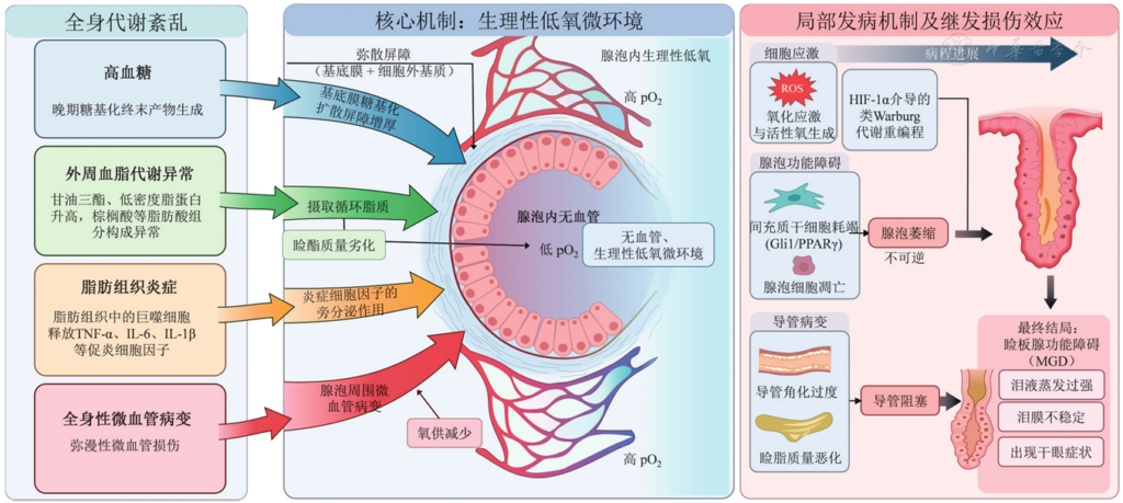
Figure 1 Pathophysiological mechanism by which the physiological hypoxic microenvironment of meibomian gland acini mediates systemic metabolic disorder–induced meibomian gland dysfunction TNF-α: tumor necrosis factor-α; IL: interleukin; pO₂: partial pressure of oxygen; ROS: reactive oxygen species; HIF-1α: hypoxia-inducible factor-1α; Gli1: zinc-finger transcription factor; PPARγ: peroxisome proliferator-activated receptor-γ
References
[1] 亚洲干眼协会中国分会, 海峡两岸医药卫生交流协会眼科学专业委员会眼表与泪液病学组, 中国医师协会眼科医师分会眼表与干眼学组. 中国睑板腺功能障碍专家共识: 定义和分类(2023年)[J]. 中华眼科杂志, 2023, 59(4):256-261. DOI: 10.3760/cma.j.cn112142-20230114-00023. Chinese Branch of the Asian Dry Eye Society, Ocular Surface and Tear Film Diseases Group of Ophthalmology Committee of Cross-Straits Medicine Exchange Association, Ocular Surface and Dry Eye Group of Chinese Ophthalmologist Association. Chinese expert consensus on meibomian gland dysfunction: definition and classification (2023)[J]. Chin J Ophthalmol, 2023, 59(4):256-261. DOI: 10.3760/cma.j.cn112142-20230114-00023.▼click to see more references
[2] Nichols KK, Foulks GN, Bron AJ, et al. The international workshop on meibomian gland dysfunction: executive summary[J]. Invest Ophthalmol Vis Sci, 2011, 52(4):1922-1929. DOI: 10.1167/iovs.10-6997a.
[3] Knop E, Knop N, Millar T, et al. The international workshop on meibomian gland dysfunction: report of the subcommittee on anatomy, physiology, and pathophysiology of the meibomian gland[J]. Invest Ophthalmol Vis Sci, 2011, 52(4):1938-1978. DOI: 10.1167/iovs.10-6997c.
[4] Graham AD, Yeh CH, Lin MC. Morphological characteristics of the meibomian glands: impact of demographics and contact lens wear[J]. Transl Vis Sci Technol, 2025, 14(8):36. DOI: 10.1167/tvst.14.8.36.
[5] Karara AM, El-Sanabary Z, El-Helw MA, et al. Standardization of meibomian gland dysfunction in an Egyptian population sample using a non-contact meibography technique[J]. Int J Ophthalmol, 2024, 17(1):61-65. DOI: 10.18240/ijo.2024.01.08.
[6] Hassanzadeh S, Varmaghani M, Zarei-Ghanavati S, et al. Global prevalence of meibomian gland dysfunction: a systematic review and meta-analysis[J]. Ocul Immunol Inflamm, 2021, 29(1):66-75. DOI: 10.1080/09273948.2020.1755441.
[7] Mussi N, Haque W, Robertson DM. The association between risk factors for metabolic syndrome and meibomian gland disease in a dry eye cohort[J]. Clin Ophthalmol, 2021, 15:3821-3832. DOI: 10.2147/OPTH.S322461.
[8] Akowuah PK, Owusu E, Senanu EN, et al. Association between dyslipidemia and meibomian gland dysfunction: a systematic review and meta-analysis[J]. Optom Vis Sci, 2023, 100(3):211-217. DOI: 10.1097/OPX.0000000000001994.
[9] Zhao X, An X, Yang C, et al. The crucial role and mechanism of insulin resistance in metabolic disease[J]. Front Endocrinol (Lausanne), 2023, 14:1149239. DOI: 10.3389/fendo.2023.1149239.
[10] Saadati S, Godini R, Reddy A, et al. Metabolic crossroads in insulin resistance: exploring lipid dysregulation and inflammation[J]. Front Immunol, 2025, 16:1692742. DOI: 10.3389/fimmu.2025.1692742.
[11] Lee YS, Olefsky J. Chronic tissue inflammation and metabolic disease[J]. Genes Dev, 2021, 35(5-6):307-328. DOI: 10.1101/gad.346312.120.
[12] Magliano DJ, Boyko EJ. IDF diabetes atlas[M]. 11th ed. Brussels: International Diabetes Federation, 2025:42.
[13] Pan C, Cao B, Fang H, et al. Global burden of diabetes mellitus 1990 2021: epidemiological trends, geospatial disparities, and risk factor dynamics[J]. Front Endocrinol (Lausanne), 2025, 16:1596127. DOI: 10.3389/fendo.2025.1596127.
[14] Viso E, Rodríguez-Ares MT, Abelenda D, et al. Prevalence of asymptomatic and symptomatic meibomian gland dysfunction in the general population of Spain[J]. Invest Ophthalmol Vis Sci, 2012, 53(6):2601-2606. DOI: 10.1167/iovs.11-9228.
[15] Schaumberg DA, Nichols JJ, Papas EB, et al. The international workshop on meibomian gland dysfunction: report of the subcommittee on the epidemiology of, and associated risk factors for, MGD[J]. Invest Ophthalmol Vis Sci, 2011, 52(4):1994-2005. DOI: 10.1167/iovs.10-6997e.
[16] Lin X, Xu B, Zheng Y, et al. Meibomian gland dysfunction in type 2 diabetic patients[J]. J Ophthalmol, 2017, 2017:3047867. DOI: 10.1155/2017/3047867.
[17] 胡皎月, 廖怿, 黄彩虹, 等. 中国成人睑板腺功能障碍与体质指数的相关性[J]. 中华医学杂志, 2021, 101(32):2514-2518. DOI: 10.3760/cma.j.cn112137-20210223-00466. Hu JY, Liao Y, Huang CH, et al. Association between meibomian gland dysfunction and body mass index in Chinese adults[J]. Natl Med J China, 2021, 101(32):2514-2518. DOI: 10.3760/cma.j.cn112137-20210223-00466.
[18] Liu Y, Chen D, Chen X, et al. Hypoxia: a breath of fresh air for the meibomian gland[J]. Ocul Surf, 2019, 17(2):310-317. DOI: 10.1016/j.jtos.2018.12.001.
[19] Liu Y, Butovich IA, Garreis F, et al. Comparative characterization of human meibomian glands, free sebaceous glands, and hair-associated sebaceous glands based on biomarkers, analysis of secretion composition, and gland morphology[J]. Int J Mol Sci, 2024, 25(6):3109. DOI: 10.3390/ijms25063109.
[20] Zhu X, Xu M, Portal C, et al. Identification of meibomian gland stem cell populations and mechanisms of aging[J]. Nat Commun, 2025, 16(1):1663. DOI: 10.1038/s41467-025-56907-6.
[21] Green-Church KB, Butovich I, Willcox M, et al. The international workshop on meibomian gland dysfunction: report of the subcommittee on tear film lipids and lipid-protein interactions in health and disease[J]. Invest Ophthalmol Vis Sci, 2011, 52(4):1979-1993. DOI: 10.1167/iovs.10-6997d.
[22] Butovich IA, Bhat N, Wojtowicz JC. Comparative transcriptomic and lipidomic analyses of human male and female meibomian glands reveal common signature genes of meibogenesis[J]. Int J Mol Sci, 2019, 20(18):4539. DOI: 10.3390/ijms20184539.
[23] Butovich IA, McMahon A, Wojtowicz JC, et al. Effects of sex (or lack thereof) on meibogenesis in mice (Mus musculus): comparative evaluation of lipidomes and transcriptomes of male and female tarsal plates[J]. Ocul Surf, 2019, 17(4):793-808. DOI: 10.1016/j.jtos.2019.03.002.
[24] Liu Y, Wang J, Chen D, et al. The role of hypoxia-inducible factor 1α in the regulation of human meibomian gland epithelial cells[J]. Invest Ophthalmol Vis Sci, 2020, 61(3):1. DOI: 10.1167/iovs.61.3.1.
[25] Cobb LM, Nolan J, O’Neill P. Microscopic distribution of misonidazole in mouse tissues[J]. Br J Cancer, 1989, 59(1):12-16. DOI: 10.1038/bjc.1989.4.
[26] Pias SC. Pathways of oxygen diffusion in cells and tissues: hydrophobic channeling via networked lipids[J]. Adv Exp Med Biol, 2020, 1232:183-190. DOI: 10.1007/978-3-030-34461-0_23.
[27] Kierans SJ, Taylor CT. Regulation of glycolysis by the hypoxia-inducible factor (HIF): implications for cellular physiology[J]. J Physiol, 2021, 599(1):23-37. DOI: 10.1113/JP280572.
[28] Yang P, Feng J, Peng Q, et al. Advanced glycation end products: potential mechanism and therapeutic target in cardiovascular complications under diabetes[J]. Oxid Med Cell Longev, 2019, 2019:9570616. DOI: 10.1155/2019/9570616.
[29] Oshitari T. Advanced glycation end-products and diabetic neuropathy of the retina[J]. Int J Mol Sci, 2023, 24(3):2927. DOI: 10.3390/ijms24032927.
[30] Roy S, Kim D. Retinal capillary basement membrane thickening: role in the pathogenesis of diabetic retinopathy[J]. Prog Retin Eye Res, 2021, 82:100903. DOI: 10.1016/j.preteyeres.2020.100903.
[31] Wu H, Fang X, Luo S, et al. Meibomian glands and tear film findings in type 2 diabetic patients: a cross-sectional study[J]. Front Med (Lausanne), 2022, 9:762493. DOI: 10.3389/fmed.2022.762493.
[32] Butovich IA, Suzuki T. Delineating a novel metabolic high triglycerides-low waxes syndrome that affects lipid homeostasis in meibomian and sebaceous glands[J]. Exp Eye Res, 2020, 199:108189. DOI: 10.1016/j.exer.2020.108189.
[33] Georgiev GA, Borchman D, Eftimov P, et al. Lipid saturation and the rheology of human tear lipids[J]. Int J Mol Sci, 2019, 20(14):3431. DOI: 10.3390/ijms20143431.
[34] Nishimura S, Manabe I, Nagai R. Adipose tissue inflammation in obesity and metabolic syndrome[J]. Discov Med, 2009, 8(41):55-60.
[35] Appari M, Channon KM, McNeill E. Metabolic regulation of adipose tissue macrophage function in obesity and diabetes[J]. Antioxid Redox Signal, 2018, 29(3):297-312. DOI: 10.1089/ars.2017.7060.
[36] Bu J, Zhang M, Wu Y, et al. High-fat diet induces inflammation of meibomian gland[J]. Invest Ophthalmol Vis Sci, 2021, 62(10):13. DOI: 10.1167/iovs.62.10.13.
[37] Jan YK, Kelhofer N, Tu T, et al. Diagnosis, pathophysiology and management of microvascular dysfunction in diabetes mellitus[J]. Diagnostics (Basel), 2024, 14(24):2830. DOI: 10.3390/diagnostics14242830.
[38] Yang X, Reneker LW, Zhong X, et al. Meibomian gland stem/progenitor cells: the hunt for gland renewal[J]. Ocul Surf, 2023, 29:497-507. DOI: 10.1016/j.jtos.2023.07.004.
[39] Zou Z, Wang H, Zhang B, et al. Inhibition of Gli1 suppressed hyperglycemia-induced meibomian gland dysfunction by promoting pparγ expression[J]. Biomed Pharmacother, 2022, 151:113109. DOI: 10.1016/j.biopha.2022.113109.