Authors:Wang Gongyue,Zhang Yijia,Zhang Mingchang,Xie Huatao
Corresponding author: Xie Huatao, Email:huataoxie@hust.edu.cn
Published:2026-10-10
DOI:10.3760/cma.j.cn115989-20260309-00098
ABSTRACT
Meibomian gland dysfunction is recognized as the primary cause of dry eye. As a lipid-secreting gland, the normal renewal and secretory function are essential for maintaining tear film stability and ocular surface homeostasis. Findings from lineage tracing, single-cell transcriptomics, and organoid models suggest that several spatially distinct stem/progenitor cell populations may be involved in maintaining meibomian gland homeostasis, which are regulated by the microenvironment, aging, and inflammation. This review summarizes current evidence regarding the localization and markers of stem/progenitor cells, the roles of the microenvironment and key signaling pathways in regulating their self-renewal and differentiation. We also discuss recent advances in meibomian gland organoid models for drug screening and targeted therapeutic development. These findings may help provide a theoretical basis and translational pathways for developing regenerative and therapeutic strategies targeting meibomian gland stem/progenitor cells.
KEYWORDS:
Meibomian gland dysfunction;Meibomian gland stem/progenitor cells;Lineage tracing;Organoids
COPYRIGHTS:
Copyright by Chinese Medical Association
No content published by the journals of Chinese Medical Association may be reproduced or abridged without authorization. Please do not use or copy the layout and design of the journals without permission.
All articles published represent the opinions of the authors, and do not reflect the official policy of the Chinese Medical Association or the Editorial Board, unless this is clearly specified.
Authors Info & Affiliations
Wang Gongyue
Department of Ophthalmology, Union Hospital, Tongji Medical College, Huazhong University of Science and Technology, Wuhan 430022, China
Zhang Yijia
Department of Ophthalmology, Union Hospital, Tongji Medical College, Huazhong University of Science and Technology, Wuhan 430022, China
Zhang Mingchang
Department of Ophthalmology, Union Hospital, Tongji Medical College, Huazhong University of Science and Technology, Wuhan 430022, China
Xie Huatao
Department of Ophthalmology, Union Hospital, Tongji Medical College, Huazhong University of Science and Technology, Wuhan 430022, China
Figures & Tables
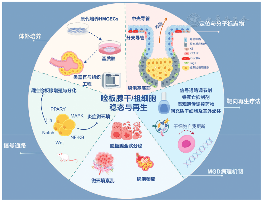
Figure 1 Homeostasis and regeneration of meibomian gland stem/progenitor cells HMGECs, human meibomian gland epithelial cells; MGD, meibomian gland dysfunction
▼click to see the rest
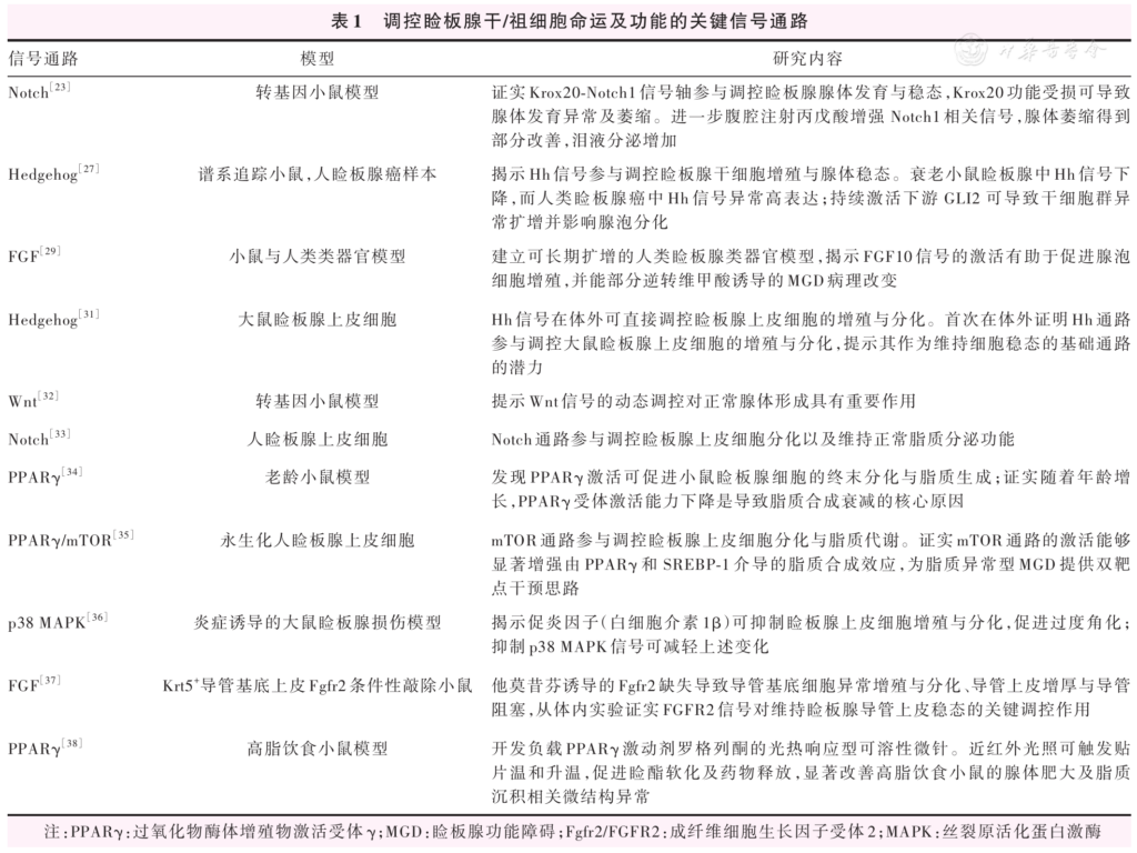
Table 1 Key signaling pathways regulating the fate and function of meibomian gland stem/progenitor cells PPARγ, peroxisome proliferator-activated receptor γ; MGD, meibomian gland dysfunction; Fgfr2/FGFR2, fibroblast growth factor receptor 2; MAPK, mitogen-activated protein kinase
References
[1] Schaumberg DA, Nichols JJ, Papas EB, et al. The international workshop on meibomian gland dysfunction: report of the subcommittee on the epidemiology of, and associated risk factors for, MGD[J]. Invest Ophthalmol Vis Sci, 2011, 52(4):1994-2005. DOI: 10.1167/iovs.10-6997e.
▼click to see more references
[2] 亚洲干眼协会中国分会, 海峡两岸医药卫生交流协会眼科学专业委员会眼表与泪液病学组, 中国医师协会眼科医师分会眼表与干眼学组. 中国睑板腺功能障碍专家共识: 诊断和治疗(2023年)[J]. 中华眼科杂志, 2023, 59(11):880-887. DOI: 10.3760/cma.j.cn112142-20230822-00054. Chinese Branch of the Asian Dry Eye Society, Ocular Surface and Tear Film Diseases Group of Ophthalmology Committee of Cross-Straits Medicine Exchange Association, Ocular Surface and Dry Eye Group of Chinese Ophthalmologist Association. Chinese expert consensus on meibomian gland dysfunction: diagnosis and management (2023)[J]. Chin J Ophthalmol, 2023, 59(11):880-887. DOI: 10.3760/cma.j.cn112142-20230822-00054.
[3] Teo C, Ong HS, Liu YC, et al. Meibomian gland dysfunction is the primary determinant of dry eye symptoms: analysis of 2346 patients[J]. Ocul Surf, 2020, 18(4):604-612. DOI: 10.1016/j.jtos.2020.06.008.
[4] Nelson JD, Shimazaki J, Benitez-del-Castillo JM, et al. The international workshop on meibomian gland dysfunction: report of the definition and classification subcommittee[J]. Invest Ophthalmol Vis Sci, 2011, 52(4):1930-1937. DOI: 10.1167/iovs.10-6997b.
[5] Craig JP, Nichols KK, Akpek EK, et al. TFOS DEWS II definition and classification report[J]. Ocul Surf, 2017, 15(3):276-283. DOI: 10.1016/j.jtos.2017.05.008.[6] Kim W, Kim DH, Eom Y, et al. Rethinking the pathogenesis of meibomian gland dysfunction: ductal-centric vs. meibocyte-centric hypothesis[J]. Exp Eye Res, 2025, 261:110646. DOI: 10.1016/j.exer.2025.110646.
[7] Knop E, Knop N, Millar T, et al. The international workshop on meibomian gland dysfunction: report of the subcommittee on anatomy, physiology, and pathophysiology of the meibomian gland[J]. Invest Ophthalmol Vis Sci, 2011, 52(4):1938-1978. DOI: 10.1167/iovs.10-6997c.
[8] Willcox M, Argüeso P, Georgiev GA, et al. TFOS DEWS II tear film report[J]. Ocul Surf, 2017, 15(3):366-403. DOI: 10.1016/j.jtos.2017.03.006.
[9] Wolffsohn JS, Benítez-Del-Castillo JM, Loya-Garcia D, et al. TFOS DEWS III: diagnostic methodology[J]. Am J Ophthalmol, 2025, 279:387-450. DOI: 10.1016/j.ajo.2025.05.033.
[10] Yang X, Reneker LW, Zhong X, et al. Meibomian gland stem/progenitor cells: the hunt for gland renewal[J]. Ocul Surf, 2023, 29:497-507. DOI: 10.1016/j.jtos.2023.07.004.
[11] Olami Y, Zajicek G, Cogan M, et al. Turnover and migration of meibomian gland cells in rats' eyelids[J]. Ophthalmic Res, 2001, 33(3):170-175. DOI: 10.1159/000055665.
[12] Hwang HS, Parfitt GJ, Brown DJ, et al. Meibocyte differentiation and renewal: insights into novel mechanisms of meibomian gland dysfunction (MGD)[J]. Exp Eye Res, 2017, 163:37-45. DOI: 10.1016/j.exer.2017.02.008.
[13] Nichols KK, Foulks GN, Bron AJ, et al. The international workshop on meibomian gland dysfunction: executive summary[J]. Invest Ophthalmol Vis Sci, 2011, 52(4):1922-1929. DOI: 10.1167/iovs.10-6997a.
[14] Wang S, Li Y, Gao J, et al. Atrophic meibomian gland dysfunction induced by eyelid margin cryotherapy with liquid nitrogen[J]. Sci Rep, 2025, 15(1):754. DOI: 10.1038/s41598-024-84742-0.
[15] Jester JV, Parfitt GJ, Brown DJ. Meibomian gland dysfunction: hyperkeratinization or atrophy?[J]. BMC Ophthalmol, 2015, 15 Suppl 1:156. DOI: 10.1186/s12886-015-0132-x.
[16] Du YL, Peng X, Liu Y, et al. Ductal hyperkeratinization and acinar renewal abnormality: new concepts on pathogenesis of meibomian gland dysfunction[J]. Curr Issues Mol Biol, 2023, 45(3):1889-1901. DOI: 10.3390/cimb45030122.
[17] Reyes NJ, Yu C, Mathew R, et al. Neutrophils cause obstruction of eyelid sebaceous glands in inflammatory eye disease in mice[J]. Sci Transl Med, 2018, 10(451):eaas9164. DOI: 10.1126/scitranslmed.aas9164.
[18] Beatty CJ, Ruiz-Lozano RE, Quiroga-Garza ME, et al. The Yin and Yang of non-immune and immune responses in meibomian gland dysfunction[J]. Ocul Surf, 2024, 32:81-90. DOI: 10.1016/j.jtos.2024.01.004.
[19] Chen C, Liao Y, Peng G. Connecting past and present: single-cell lineage tracing[J]. Protein Cell, 2022, 13(11):790-807. DOI: 10.1007/s13238-022-00913-7.
[20] Short S, García-Tejera R, Schumacher LJ, et al. Next generation lineage tracing and its applications to unravel development[J]. NPJ Syst Biol Appl, 2025, 11(1):60. DOI: 10.1038/s41540-025-00542-w.
[21] Parfitt GJ, Geyfman M, Xie Y, et al. Characterization of quiescent epithelial cells in mouse meibomian glands and hair follicle/sebaceous glands by immunofluorescence tomography[J]. J Invest Dermatol, 2015, 135(4):1175-1177. DOI: 10.1038/jid.2014.484.
[22] Parfitt GJ, Lewis PN, Young RD, et al. Renewal of the holocrine meibomian glands by label-retaining, unipotent epithelial progenitors[J]. Stem Cell Reports, 2016, 7(3):399-410. DOI: 10.1016/j.stemcr.2016.07.010.
[23] Zhang Y, Tchegnon E, Ghotbi E, et al. Disruption of Krox20-Notch1 signaling blocks meibomian gland development and homeostasis leading to dry eye disease[J]. Nat Commun, 2025, 16(1):11584. DOI: 10.1038/s41467-025-66740-6.
[24] Tchegnon E, Liao CP, Ghotbi E, et al. Epithelial stem cell homeostasis in meibomian gland development, dysfunction, and dry eye disease[J]. JCI Insight, 2021, 6(20):e151078. DOI: 10.1172/jci.insight.151078.
[25] Zhu X, Xu M, Owens DM, et al. KRT6A and KRT17 mark distinct stem cell populations in the adult palpebral conjunctiva and meibomian gland[J]. Cells, 2025, 14(24):1979. DOI: 10.3390/cells14241979.
[26] Guo Y, Zhang R, Zhang M, et al. Assessment of the clonal growth potential of meibomian gland stem/progenitor cells via clonal analysis[J]. Ocul Surf, 2025, 37:1-10. DOI: 10.1016/j.jtos.2025.02.006.
[27] Zhu X, Xu M, Portal C, et al. Identification of meibomian gland stem cell populations and mechanisms of aging[J]. Nat Commun, 2025, 16(1):1663. DOI: 10.1038/s41467-025-56907-6.
[28] Xie HT, Sullivan DA, Chen D, et al. Biomarkers for progenitor and differentiated epithelial cells in the human meibomian gland[J]. Stem Cells Transl Med, 2018, 7(12):887-892. DOI: 10.1002/sctm.18-0037.
[29] Yu C, Wan X, Wei J, et al. Human meibomian gland organoids to study epithelial homeostasis and dysfunction[J]. Protein Cell, 2026, 17(8):775-789. DOI: 10.1093/procel/pwaf095.
[30] Veniaminova NA, Jia YY, Hartigan AM, et al. Distinct mechanisms for sebaceous gland self-renewal and regeneration provide durability in response to injury[J]. Cell Rep, 2023, 42(9):113121. DOI: 10.1016/j.celrep.2023.113121.
[31] Qu JY, Xiao YT, Zhang YY, et al. Hedgehog signaling pathway regulates the proliferation and differentiation of rat meibomian gland epithelial cells[J]. Invest Ophthalmol Vis Sci, 2021, 62(2):33. DOI: 10.1167/iovs.62.2.33.
[32] Sima J, Piao Y, Chen Y, et al. Molecular dynamics of Dkk4 modulates Wnt action and regulates meibomian gland development[J]. Development, 2016, 143(24):4723-4735. DOI: 10.1242/dev.143909.
[33] Gidfar S, Afsharkhamseh N, Sanjari S, et al. Notch signaling in meibomian gland epithelial cell differentiation[J]. Invest Ophthalmol Vis Sci, 2016, 57(3):859-865. DOI: 10.1167/iovs.15-18319.
[34] Jester JV, Potma E, Brown DJ. PPARγ regulates mouse meibocyte differentiation and lipid synthesis[J]. Ocul Surf, 2016, 14(4):484-494. DOI: 10.1016/j.jtos.2016.08.001.
[35] Jun I, Choi YJ, Kim BR, et al. Activation of the mTOR pathway enhances PPARγ/SREBP-mediated lipid synthesis in human meibomian gland epithelial cells[J]. Sci Rep, 2024, 14(1):28118. DOI: 10.1038/s41598-024-73969-6.
[36] Qu JY, Xie HT, Xiao YT, et al. The inhibition of p38 MAPK blocked inflammation to restore the functions of rat meibomian gland epithelial cells[J]. Exp Eye Res, 2023, 231:109470. DOI: 10.1016/j.exer.2023.109470.
[37] Yang X, Zhong X, Lin H, et al. Deletion of Fgfr2 in ductal basal epithelium with tamoxifen induces obstructive meibomian gland dysfunction[J]. Invest Ophthalmol Vis Sci, 2024, 65(13):36. DOI: 10.1167/iovs.65.13.36.
[38] Yu F, Zhao X, Wang Q, et al. Photothermal-responsive soluble microneedle patches for meibomian gland dysfunction therapy[J]. Adv Sci (Weinh), 2025, 12(11):e2413962. DOI: 10.1002/advs.202413962.
[39] Chen H, Gao H, Xie HT, et al. Hyperkeratinization and proinflammatory cytokine expression in meibomian glands induced by staphylococcus aureus[J]. Invest Ophthalmol Vis Sci, 2021, 62(13):11. DOI: 10.1167/iovs.62.13.11.
[40] Moreno I, Verma S, Gesteira TF, et al. Recent advances in age-related meibomian gland dysfunction (ARMGD)[J]. Ocul Surf, 2023, 30:298-306. DOI: 10.1016/j.jtos.2023.11.003.
[41] Wang H, Zou Z, Wan L, et al. Periplocin ameliorates mouse age-related meibomian gland dysfunction through up-regulation of Na/K-ATPase via SRC pathway[J]. Biomed Pharmacother, 2022, 146:112487. DOI: 10.1016/j.biopha.2021.112487.
[42] Wu R, Liu S, Shen Z, et al. Corneal oxidative stress, inflammation, and senescence as key drivers of ocular pathology in a chronic graft-versus-host disease model[J]. BMC Ophthalmol, 2025, 25(1):379. DOI: 10.1186/s12886-025-04210-9.
[43] Yoon CH, Ryu JS, Hwang HS, et al. Comparative analysis of age-related changes in lacrimal glands and meibomian glands of a C57BL/6 male mouse model[J]. Int J Mol Sci, 2020, 21(11):4169. DOI: 10.3390/ijms21114169.
[44] Verma S, Moreno IY, Sun M, et al. Age related changes in hyaluronan expression leads to meibomian gland dysfunction[J]. Matrix Biol, 2023, 124:23-38. DOI: 10.1016/j.matbio.2023.11.002.
[45] Zhu X, Xu M, Millar SE. HDAC1/2 and HDAC3 play distinct roles in controlling adult Meibomian gland homeostasis[J]. Ocul Surf, 2024, 33:39-49. DOI: 10.1016/j.jtos.2024.04.005.
[46] Zhu X, Huang S. Epigenetic regulation of sebaceous and meibomian glands: from development to disease[J]. Biomedicines, 2026, 14(2):468. DOI: 10.3390/biomedicines14020468.
[47] Liu S, Hatton MP, Khandelwal P, et al. Culture, immortalization, and characterization of human meibomian gland epithelial cells[J]. Invest Ophthalmol Vis Sci, 2010, 51(8):3993-4005. DOI: 10.1167/iovs.09-5108.
[48] Phan M, Madigan MC, Stapleton F, et al. Human meibomian gland epithelial cell culture models: current progress, challenges, and future directions[J]. Ocul Surf, 2022, 23:96-113. DOI: 10.1016/j.jtos.2021.11.012.
[49] Peng X, Du YL, Liu ST, et al. Isolation and culture of human meibomian gland ductal cells[J]. Invest Ophthalmol Vis Sci, 2023, 64(15):29. DOI: 10.1167/iovs.64.15.29.
[50] Zahn I, Garreis F, Schicht M, et al. A new organotypic 3D slice culture of mouse meibomian glands reveals impact of melanocortins[J]. Int J Mol Sci, 2022, 23(23):14947. DOI: 10.3390/ijms232314947.
[51] Wang G, Xu L, Shi R, et al. Organotypic culture model of mouse meibomian gland as a screening platform for risk factors related to meibomian gland dysfunction[J]. Ocul Surf, 2023, 30:73-84. DOI: 10.1016/j.jtos.2023.08.007.
[52] Asano N, Hampel U, Garreis F, et al. Differentiation patterns of immortalized human meibomian gland epithelial cells in three-dimensional culture[J]. Invest Ophthalmol Vis Sci, 2018, 59(3):1343-1353. DOI: 10.1167/iovs.17-23266.
[53] Sun M, Puri S, Parfitt GJ, et al. Hyaluronan regulates eyelid and meibomian gland morphogenesis[J]. Invest Ophthalmol Vis Sci, 2018, 59(8):3713-3727. DOI: 10.1167/iovs.18-24292.
[54] Xu KK, Huang YK, Liu X, et al. Organotypic culture of mouse meibomian gland: a novel model to study meibomian gland dysfunction in vitro[J]. Invest Ophthalmol Vis Sci, 2020, 61(4):30. DOI: 10.1167/iovs.61.4.30.
[55] Nuwormegbe S, Park NY, Park HJ, et al. Induction of meibocyte differentiation by three-dimensional, matrigel culture of immortalized human meibomian gland epithelial cells to form acinar organoids[J]. Ocul Surf, 2022, 26:271-282. DOI: 10.1016/j.jtos.2022.10.004.
[56] Qu J, Yang L, Wang X, et al. Establishment of mouse meibomian gland organoids for in vitro modeling of meibomian gland dysfunction[J]. Invest Ophthalmol Vis Sci, 2026, 67(1):6. DOI: 10.1167/iovs.67.1.6.
[57] Chen L, Yan D, Wu N, et al. 3D-printed poly-caprolactone scaffolds modified with biomimetic extracellular matrices for tarsal plate tissue engineering[J]. Front Bioeng Biotechnol, 2020, 8:219. DOI: 10.3389/fbioe.2020.00219.
[58] Lee H, Park YH, Kang HJ, et al. Three-dimensional bioprinting of tarsal plates with adipose-derived mesenchymal stem cells: evaluation of meibomian gland reconstruction in a rat model[J]. Biomedicines, 2024, 12(11):2567. DOI: 10.3390/biomedicines12112567.
[59] Bu J, Guo Y, Wu Y, et al. Models for meibomian gland dysfunction: in vivo and in vitro[J]. Ocul Surf, 2024, 32:154-165. DOI: 10.1016/j.jtos.2024.03.003.
[60] 黎颖莉, 刘祖国. 睑板腺功能障碍药物治疗研究进展[J]. 中华实验眼科杂志, 2023, 41(5):493-498. DOI: 10.3760/cma.j.cn115989-20211202-00668. Li YL, Liu ZG. Advances in drug therapy of meibomian gland dysfunction[J]. Chin J Exp Ophthalmol, 2023, 41(5):493-498. DOI: 10.3760/cma.j.cn115989-20211202-00668.
[61] Luo S, Guo Y, Liu Z. Current pharmaceutical therapies for meibomian gland dysfunction: from basic research to clinical practice[J]. Clin Exp Ophthalmol, 2025, 53(8):1039-1055. DOI: 10.1111/ceo.14577.
[62] Wu M, He HL, Wang C, et al. Maresin 1 ameliorates blue light overexposure-induced meibomian gland dysfunction via inhibition of NLRP3/Caspase-1/GSDMD-mediated pyroptosis[J]. Invest Ophthalmol Vis Sci, 2025, 66(13):29. DOI: 10.1167/iovs.66.13.29.
[63] Liu R, Li J, Xu Y, et al. Melatonin attenuates lps-induced proinflammatory cytokine response and lipogenesis in human meibomian gland epithelial cells via MAPK/NF-κB pathway[J]. Invest Ophthalmol Vis Sci, 2022, 63(5):6. DOI: 10.1167/iovs.63.5.6.
[64] Guo Y, Luo S, Li W, et al. Ferroptosis in meibomian gland progenitor cells contributes to pathogenesis of meibomian gland dysfunction[J]. Invest Ophthalmol Vis Sci, 2026, 67(1):9. DOI: 10.1167/iovs.67.1.9.
[65] Ko EK, Anderson A, D'souza C, et al. Disruption of H3K36 methylation provokes cellular plasticity to drive aberrant glandular formation and squamous carcinogenesis[J]. Dev Cell, 2024, 59(2):187-198. DOI: 10.1016/j.devcel.2023.12.007.
[66] Wang W, Wang H, Liu X, et al. E2F2(E2F transcription factor 2) as a potential therapeutic target in meibomian gland carcinoma: evidence from functional and epigenetic studies[J]. BMC Cancer, 2025, 25(1):880. DOI: 10.1186/s12885-025-13833-6.
[67] Kam WR, Sullivan DA. Neurotransmitter influence on human meibomian gland epithelial cells[J]. Invest Ophthalmol Vis Sci, 2011, 52(12):8543-8548. DOI: 10.1167/iovs.11-8113.
[68] Fan NW, Ho TC, Lin EH, et al. Pigment epithelium-derived factor peptide reverses mouse age-related meibomian gland atrophy[J]. Exp Eye Res, 2019, 185:107678. DOI: 10.1016/j.exer.2019.05.018.
[69] Thode AR, Latkany RA. Current and emerging therapeutic strategies for the treatment of meibomian gland dysfunction (MGD)[J]. Drugs, 2015, 75(11):1177-1185. DOI: 10.1007/s40265-015-0432-8.
[70] Li YZ, Wang C, Peng X, et al. Caffeine's protective role in dry eye disease and meibomian gland dysfunction: insights from clinical and experimental models[J]. Int Immunopharmacol, 2025, 146:113863. DOI: 10.1016/j.intimp.2024.113863.
[71] Cai Y, Zhang X, Yang C, et al. Melatonin alleviates high-fat-diet-induced dry eye by regulating macrophage polarization via IFT27 and lowering ERK/JNK phosphorylation[J]. iScience, 2024, 27(7):110367. DOI: 10.1016/j.isci.2024.110367.
[72] Wang C, Li YZ, Guo H, et al. Melatonin alleviates age-related lacrimal gland dysfunction via SIRT-1/NLRP3 Pathway[J]. Invest Ophthalmol Vis Sci, 2025, 66(2):51. DOI: 10.1167/iovs.66.2.51.
[73] Beyazyıldız E, Pınarlı FA, Beyazyıldız O, et al. Efficacy of topical mesenchymal stem cell therapy in the treatment of experimental dry eye syndrome model[J]. Stem Cells Int, 2014, 2014:250230. DOI: 10.1155/2014/250230.
[74] Wang G, Li H, Long H, et al. Exosomes derived from mouse adipose-derived mesenchymal stem cells alleviate benzalkonium chloride-induced mouse dry eye model via inhibiting NLRP3 inflammasome[J]. Ophthalmic Res, 2022, 65(1):40-51. DOI: 10.1159/000519458.
[75] Møller-Hansen M, Larsen AC, Toft PB, et al. Safety and feasibility of mesenchymal stem cell therapy in patients with aqueous deficient dry eye disease[J]. Ocul Surf, 2021, 19:43-52. DOI: 10.1016/j.jtos.2020.11.013.
[76] Møller-Hansen M, Larsen AC, Wiencke AK, et al. Allogeneic mesenchymal stem cell therapy for dry eye disease in patients with Sjögren's syndrome: a randomized clinical trial[J]. Ocul Surf, 2024, 31:1-8. DOI: 10.1016/j.jtos.2023.11.007.
[77] Zhang D, Chen T, Liang Q, et al. A first-in-human, prospective pilot trial of umbilical cord-derived mesenchymal stem cell eye drops therapy for patients with refractory non-Sjögren's and Sjögren's syndrome dry eye disease[J]. Stem Cell Res Ther, 2025, 16(1):202. DOI: 10.1186/s13287-025-04292-8.
[78] Chen KY, Chan HC, Chan CM. Efficacy and safety of stem cell therapy for dry eye disease: a systematic review and meta-analysis[J]. Stem Cell Res Ther, 2026, 17(1):224. DOI: 10.1186/s13287-026-04915-8.
[79] Habibi A, Khosravi A, Soleimani M, et al. Efficacy of topical mesenchymal stem cell exosome in Sjögren's syndrome-related dry eye: a randomized clinical trial[J]. BMC Ophthalmol, 2025, 25(1):299. DOI: 10.1186/s12886-025-04078-9.