Authors:Zhou Tianyu, Huang Xiaoyu, Shen Jiachao, Zhong Hongtao, Li Guigang
Corresponding author: Li Guigang, Email:guigangli@hust.edu.cn
Published:2026-10-10
DOI:10.3760/cma.j.cn115989-20260310-00109
ABSTRACT
Objective To establish reference data for lacrimal gland size in Chinese adults using high-resolution magnetic resonance imaging (MRI), and to investigate the correlations between lacrimal gland size and demographic parameters (sex, age, height, weight, and body mass index [BMI]).
Methods A cross-sectional study was conducted. Thirty-one patients (62 eyes) who underwent brain MRI at the Department of Radiology, Tongji Hospital Affiliated to Tongji Medical College, Huazhong University of Science and Technology from August to November 2023 were enrolled. Morphological measurements of the bilateral lacrimal glands were performed using a GE Healthcare 3D/BRAVO sequence, including lacrimal gland length, maximum thickness, maximum area, and lacrimal gland volume. Patients were divided into a male group (17 patients) and a female group (14 patients) according to sex, and into a <36 years group (10 patients), a 36-<54 years group (9 patients), and a ≥54 years group (12 patients) according to age. Differences in each parameter were compared between different sexes, eyes, and age groups. Correlations among age, height, weight, BMI, and right lacrimal gland volume and left lacrimal gland volume were analyzed. This study complied with the Declaration of Helsinki. The study protocol was approved by the Ethics Committee of Tongji Hospital Affiliated to Tongji Medical College, Huazhong University of Science and Technology (No. TJ-IRB20230899). Informed consent was waived by the ethics committee owing to the retrospective nature of the study.
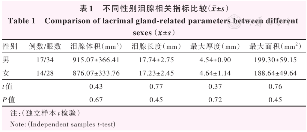
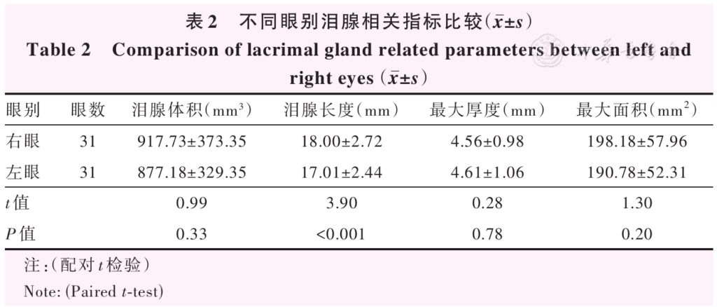
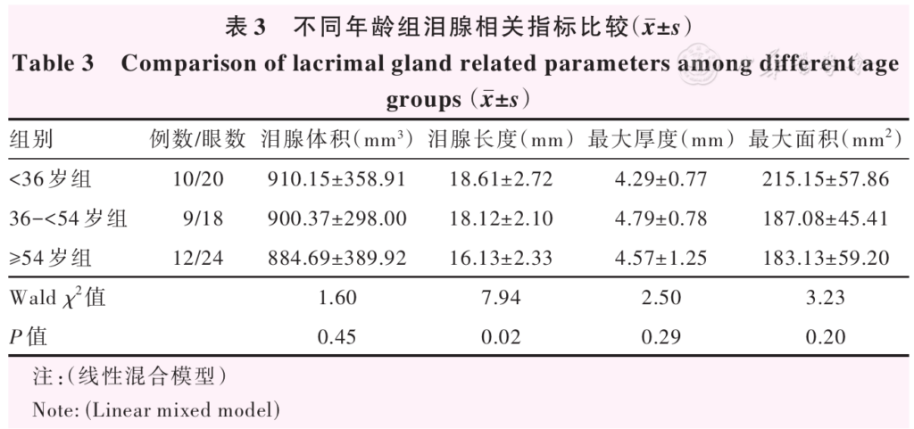
Results The mean lacrimal gland volume, lacrimal gland length, and maximum area in males were greater than those in females, while the mean maximum thickness was smaller than that in females, with no statistically significant differences (t=0.43, 0.77, 0.76, 0.37; all P>0.05). The right lacrimal gland length was (18.00±2.72)mm, which was greater than the (17.01±2.44)mm of the left lacrimal gland, and the difference was statistically significant (t=3.90, P<0.001). There were no statistically significant differences in lacrimal gland volume, maximum thickness, or maximum area between the right and left eyes (t=0.99, 0.28, 1.30; all P>0.05). The mean lacrimal gland lengths in the <36 years, 36-<54 years, and ≥54 years groups were (18.61±2.72), (18.12±2.10), and (16.13±2.33)mm, respectively, with a statistically significant overall difference (Wald χ2=7.94, P=0.02). There were no statistically significant differences in lacrimal gland volume, maximum thickness, or maximum area among the different age groups (Wald χ 2=1.60, 2.50, 3.23; all P>0.05). Correlation analysis showed that age, height, weight, and BMI were not significantly correlated with lacrimal gland volume (all P>0.05). The volumes of the left and right lacrimal glands showed a significant positive correlation (r=0.73,P<0.001).
Conclusions The right lacrimal gland long axis is significantly longer than the left one in Chinese adults, and lacrimal gland length shows a decreasing trend with age.
KEYWORDS:
Magnetic resonance imaging;Lacrimal gland volume;Lacrimal gland length;Lacrimal gland disease;Chinese adults
COPYRIGHTS:
Copyright by Chinese Medical Association
No content published by the journals of Chinese Medical Association may be reproduced or abridged without authorization. Please do not use or copy the layout and design of the journals without permission.
All articles published represent the opinions of the authors, and do not reflect the official policy of the Chinese Medical Association or the Editorial Board, unless this is clearly specified.
Authors Info & Affiliations
Zhou Tianyu
Department of Ophthalmology, Tongji Hospital Affiliated to Tongji Medical College, Huazhong University of Science and Technology, Wuhan 430030, China
Huang Xiaoyu
Department of Ophthalmology, Tongji Hospital Affiliated to Tongji Medical College, Huazhong University of Science and Technology, Wuhan 430030, China
Shen Jiachao
Department of Ophthalmology, Tongji Hospital Affiliated to Tongji Medical College, Huazhong University of Science and Technology, Wuhan 430030, China
Zhong Hongtao
Department of Radiology, Tongji Hospital Affiliated to Tongji Medical College, Huazhong University of Science and Technology, Wuhan 430030, China
Li Guigang
Department of Ophthalmology, Tongji Hospital Affiliated to Tongji Medical College, Huazhong University of Science and Technology, Wuhan 430030, China
Figures & Tables
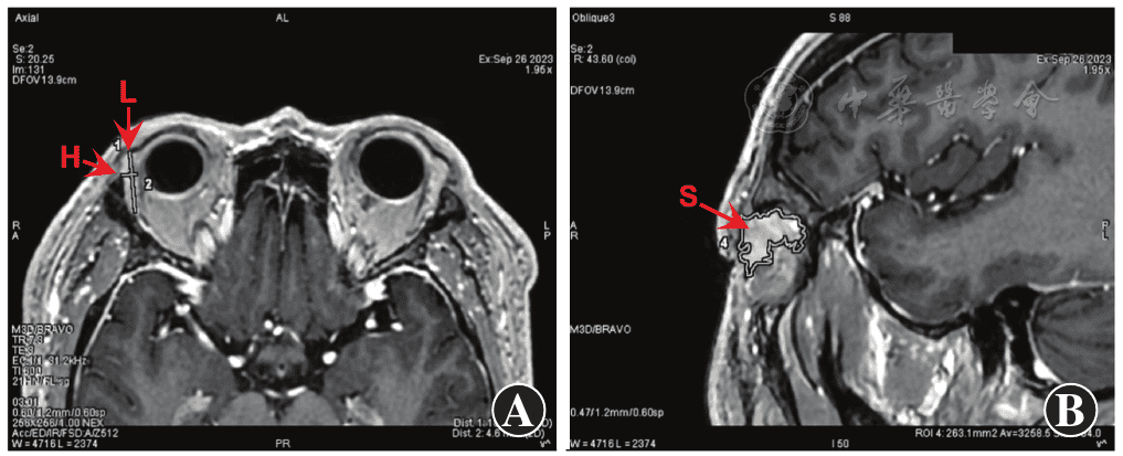
Figure 1 Lacrimal gland volume measurement A: On the human axial plane of the image, the maximum thickness (H) and maximum length (L) of the lacrimal gland were measured perpendicular to the outer surface of the eyeball B: On the rotated three-dimensional reconstruction image with the maximum length as the axis, the inclined surface of the lacrimal gland covering the largest area of the outer surface of the eyeball (S) was found and this area was calculated
▼click to see the rest
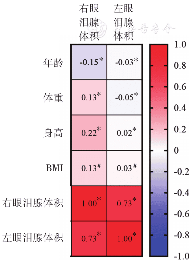
Figure 2 Heat map showing correlation between different factors and bilateral lacrimal gland volumes (*: Pearson correlation analysis; #: Spearman rank correlation analysis; n=31) The volumes of the left and right lacrimal glands showed a significant positive correlation (r=0.73,P<0.001) BMI: body mass index
References
[1] Adewoye AH, Ramsey J, McMahon L, et al. Lacrimal gland enlargement in sickle cell disease[J]. Am J Hematol, 2006, 81(11):888-889. DOI: 10.1002/ajh.20658.
▼click to see more references
[2] Rana K, Caltabiano C, Beecher M, et al. Lacrimal gland enlargement in thyroid eye disease[J]. Int Ophthalmol, 2024, 44(1):431. DOI: 10.1007/s10792-024-03352-x.
[3] Lorber M, Vidić B. Measurements of lacrimal glands from cadavers, with descriptions of typical glands and three gross variants[J]. Orbit, 2009, 28(2-3):137-146. DOI: 10.1080/01676830902766014.
[4] Karadeniz H, Cerit M, Güler AA, et al. Lacrimal gland ultrasonography and elastography as a diagnostic and activity tool for primary Sjögren's syndrome[J]. Int J Rheum Dis, 2023, 26(6):1083-1090. DOI: 10.1111/1756-185X.14702.
[5] Harris MA, Realini T, Hogg JP, et al. CT dimensions of the lacrimal gland in Graves orbitopathy[J]. Ophthalmic Plast Reconstr Surg, 2012, 28(1):69-72. DOI: 10.1097/IOP.0b013e31823c4a3a.
[6] Tenzel PA, Moffa D, Decilveo AP, et al. Normal lacrimal gland volumes by magnetic resonance imaging and the relationship of lacrimal gland volume to orbital size[J]. J Craniofac Surg, 2019, 30(8):e741-e743. DOI: 10.1097/SCS.0000000000005752.
[7] Liu X, Song J. The application of autopsy and explanted heart samples in scientific research[J]. Cardiovasc Pathol, 2022, 59:107424. DOI: 10.1016/j.carpath.2022.107424.
[8] Dietrich CF, Ignee A, Hocke M, et al. Pitfalls and artefacts using contrast enhanced ultrasound[J]. Z Gastroenterol, 2011, 49(3):350-356. DOI: 10.1055/s-0029-1245851.
[9] Patel PR, De Jesus O. CT scan[M/OL]. Treasure Island (FL):StatPearls Publishing, 2023-01-02. https://pubmed.ncbi.nlm.nih.gov/33620865/.
[10] What is an MRI scan and what can it do?[J]. Drug Ther Bull, 2011, 49(12):141-144. DOI: 10.1136/dtb.2011.02.0073.
[11] Jordan DR, Anderson RB, Mamalis N. Accessory lacrimal glands[J]. Ophthalmic Surg, 1990, 21(2):146-147.
[12] Singh S, Basu S. The human lacrimal gland: historical perspectives, current understanding, and recent advances[J]. Curr Eye Res, 2020, 45(10):1188-1198. DOI: 10.1080/02713683.2020.1774065.
[13] 葛坚, 刘奕志. 眼科手术学[M]. 3版. 北京:人民卫生出版社, 2015:111.
[14] Bingham CM, Castro A, Realini T, et al. Calculated CT volumes of lacrimal glands in normal Caucasian orbits[J]. Ophthalmic Plast Reconstr Surg, 2013, 29(3):157-159. DOI: 10.1097/IOP.0b013e3182859751.
[15] Idowu BM, Onigbinde SO, Chen AL, et al. Relationship between the computed tomographic volumetry of the eyeballs and lacrimal glands in a Nigerian population[J]. J West Afr Coll Surg, 2022, 12(1):34-40. DOI: 10.4103/jwas.jwas_66_22.
[16] Bukhari AA, Basheer NA, Joharjy HI. Age, gender, and interracial variability of normal lacrimal gland volume using MRI[J]. Ophthalmic Plast Reconstr Surg, 2014, 30(5):388-391. DOI: 10.1097/IOP.0000000000000117.
[17] Bulbul E, Yazici A, Yanik B, et al. Evaluation of lacrimal gland dimensions and volume in Turkish population with computed tomography[J]. J Clin Diagn Res, 2016, 10(2):TC06-08. DOI: 10.7860/JCDR/2016/16331.7207.
[18] Rana K, Juniat V, Patel S, et al. Normative lacrimal gland dimensions by magnetic resonance imaging in an Australian cohort[J]. Orbit, 2023, 42(2):157-160. DOI: 10.1080/01676830.2022.2055085.