Authors:Liu Wenqi, Feng Jun, Jie Ying
Corresponding author: Jie Ying, Email:jie_yingcn@aliyun.com
Published:2026-10-10
DOI:10.3760/cma.j.cn115989-20241129-00328
ABSTRACT
Accurate assessment of tear meniscus morphology is critical for the diagnosis and treatment of ocular surface diseases such as dry eye disease. It has been found that the measurement of tear meniscus parameters is affected by a variety of factors and has become more accurate with the introduction of advanced equipment such as optical coherence tomography and comprehensive ocular surface analysers. In addition, the application of artificial intelligence and deep learning technologies has opened up new possibilities for fully automated analysis of tear meniscus morphology, improving the efficiency and accuracy of measurements. This paper systematically reviews the various methods of measuring tear meniscus parameters, including height, radius, area, and volume, and the value of these parameters in the diagnosis of dry eye disease. The assessment of tear meniscus parameters will become more automated and precise, and the advancement of tear meniscus morphology assessment technology will contribute to a better understanding of the pathophysiological mechanisms of dry eye disease and promote the development of personalized therapeutic strategies, thereby improving the quality of life of patients.
KEYWORDS:
Tear meniscus morphological assessment;Tear meniscus parameters;Dynamic tear meniscus;Artificial intelligence
COPYRIGHTS:
Copyright by Chinese Medical Association
No content published by the journals of Chinese Medical Association may be reproduced or abridged without authorization. Please do not use or copy the layout and design of the journals without permission.
All articles published represent the opinions of the authors, and do not reflect the official policy of the Chinese Medical Association or the Editorial Board, unless this is clearly specified.
Authors Info & Affiliations
Liu Wenqi
Beijing Tongren Eye Center, Beijing Tongren Hospital, Capital Medical University, Beijing Institute of Ophthalmology, Beijing Key Laboratory of Ophthalmology & Visual Sciences, Beijing 100730, China
Feng Jun
Beijing Tongren Eye Center, Beijing Tongren Hospital, Capital Medical University, Beijing Institute of Ophthalmology, Beijing Key Laboratory of Ophthalmology & Visual Sciences, Beijing 100730, China
Jie Ying
Beijing Tongren Eye Center, Beijing Tongren Hospital, Capital Medical University, Beijing Institute of Ophthalmology, Beijing Key Laboratory of Ophthalmology & Visual Sciences, Beijing 100730, China
Figures & Tables
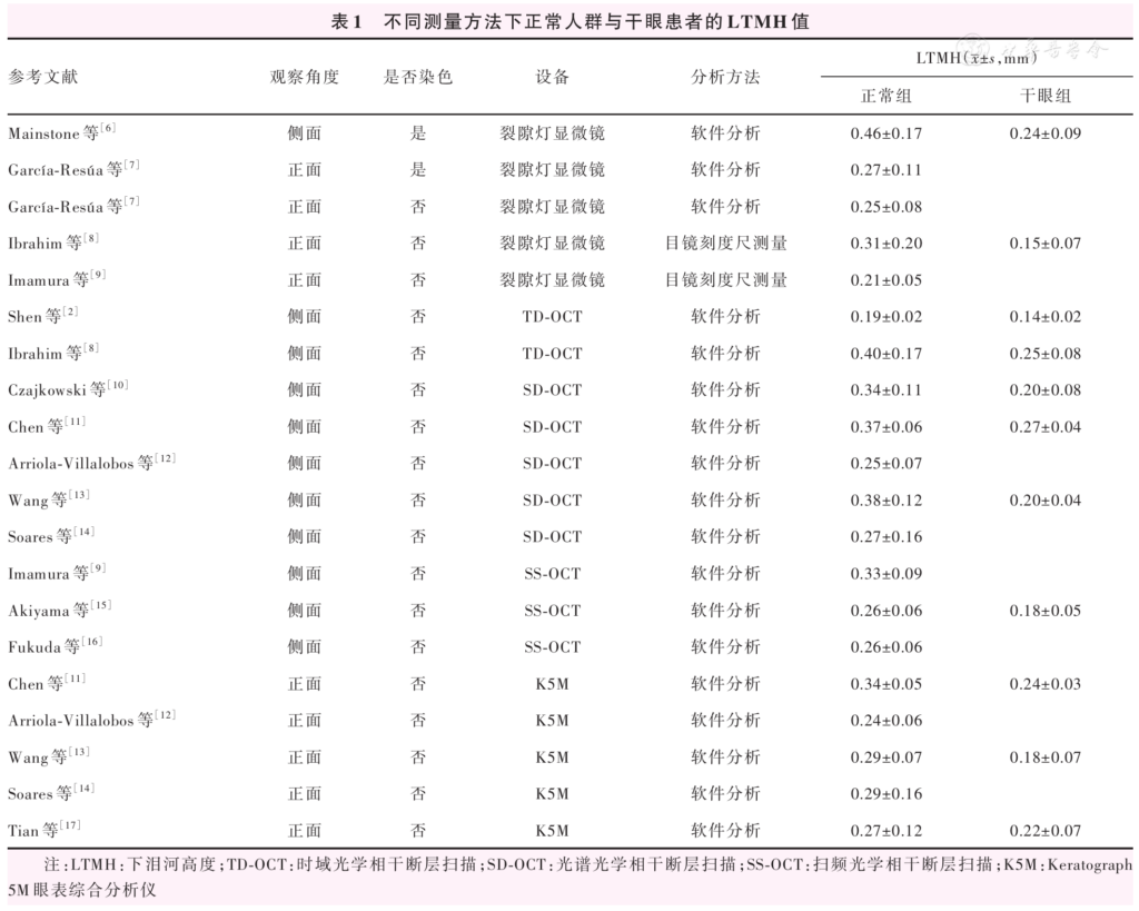
Table 1 Lower tear meniscus height (LTMH) in healthy subjects and dry eye patients measured by different methods LTMH:lower tear meniscus height; TD-OCT: time-domain optical coherence tomography; SD-OCT: spectral-domain optical coherence tomography; SS-OCT: swept-source optical coherence tomography; K5M:Keratograph 5M ocular surface analyzer
References
[1] Niedernolte B, Trunk L, Wolffsohn JS, et al. Evaluation of tear meniscus height using different clinical methods[J]. Clin Exp Optom, 2021, 104(5):583-588. DOI: 10.1080/08164622.2021.1878854.
▼click to see more references
[2] Shen M, Li J, Wang J, et al. Upper and lower tear menisci in the diagnosis of dry eye[J]. Invest Ophthalmol Vis Sci, 2009, 50(6):2722-2726. DOI: 10.1167/iovs.08-2704.
[3] 韩竹梅, 张兴儒. 结膜松弛症研究进展[J]. 国际眼科纵览, 2011, 35(6):394-399. DOI: 10.3760/cma.j.issn.1673-5803.2011.06.008.
[4] Wolffsohn JS, Benítez-Del-Castillo JM, Loya-Garcia D, et al. TFOS DEWS III: diagnostic methodology[J]. Am J Ophthalmol, 2025, 279:387-450. DOI: 10.1016/j.ajo.2025.05.033.
[5] 吴元, 吴海龙, 李海丽, 等. OCT前节模块测量的上下泪河数值与干眼的相关性[J]. 中华实验眼科杂志, 2014, 32(6):541-545. DOI: 10.3760/cma.j.issn.2095-0160.2014.06.013. Wu Y, Wu HL, Li HL, et al. Correlation between measurement of tear meniscus by anterior segment module of OCT with dry eye signs and symptoms[J]. Chin J Exp Ophthalmol, 2014, 32(6):541-545. DOI: 10.3760/cma.j.issn.2095-0160.2014.06.013.[6] Mainstone JC, Bruce AS, Golding TR. Tear meniscus measurement in the diagnosis of dry eye[J]. Curr Eye Res, 1996, 15(6):653-661. DOI: 10.3109/02713689609008906.
[7] García-Resúa C, Santodomingo-Rubido J, Lira M, et al. Clinical assessment of the lower tear meniscus height[J]. Ophthalmic Physiol Opt, 2009, 29(5):487-496. DOI: 10.1111/j.1475-1313.2009.00634.x.
[8] Ibrahim OM, Dogru M, Takano Y, et al. Application of visante optical coherence tomography tear meniscus height measurement in the diagnosis of dry eye disease[J]. Ophthalmology, 2010, 117(10):1923-1929. DOI: 10.1016/j.ophtha.2010.01.057.
[9] Imamura H, Tabuchi H, Nakakura S, et al. Usability and reproducibility of tear meniscus values generated via swept-source optical coherence tomography and the slit lamp with a graticule method[J]. Int Ophthalmol, 2018, 38(2):679-686. DOI: 10.1007/s10792-017-0517-3.
[10] Czajkowski G, Kaluzny BJ, Laudencka A, et al. Tear meniscus measurement by spectral optical coherence tomography[J]. Optom Vis Sci, 2012, 89(3):336-342. DOI: 10.1097/OPX.0b013e318242042b.
[11] Chen M, Wei A, Xu J, et al. Application of Keratograph and Fourier-domain optical coherence tomography in measurements of tear meniscus height[J]. J Clin Med, 2022, 11(5):1343. DOI: 10.3390/jcm11051343.
[12] Arriola-Villalobos P, Fernández-Vigo JI, Díaz-Valle D, et al. Assessment of lower tear meniscus measurements obtained with Keratograph and agreement with Fourier-domain optical-coherence tomography[J]. Br J Ophthalmol, 2015, 99(8):1120-1125. DOI: 10.1136/bjophthalmol-2014-306453.
[13] Wang Y, Xu Z, Gong Q, et al. The role of different tear volume detection methods in the evaluation and diagnosis of mild dry eye disease[J]. Transl Vis Sci Technol, 2022, 11(3):15. DOI: 10.1167/tvst.11.3.15.
[14] Soares I, Ramalho E, Brardo FM, et al. Tear meniscus height agreement and reproducibility between two corneal topographers and spectral-domain optical coherence tomography[J]. Clin Exp Optom, 2025, 108(4):430-436. DOI: 10.1080/08164622.2024.2341833.
[15] Akiyama R, Usui T, Yamagami S. Diagnosis of dry eye by tear meniscus measurements using anterior segment swept source optical coherence tomography[J]. Cornea, 2015, 34 Suppl 11:S115-120. DOI: 10.1097/ICO.0000000000000583.
[16] Fukuda R, Usui T, Miyai T, et al. Tear meniscus evaluation by anterior segment swept-source optical coherence tomography[J]. Am J Ophthalmol, 2013, 155(4):620-624. DOI: 10.1016/j.ajo.2012.11.009.
[17] Tian L, Qu JH, Zhang XY, et al. Repeatability and reproducibility of noninvasive Keratograph 5M measurements in patients with dry eye disease[J]. J Ophthalmol, 2016, 2016:8013621. DOI: 10.1155/2016/8013621.
[18] Lange AP, Sadjadi R, Saeedi J, et al. Time-domain and spectral-domain optical coherence tomography of retinal nerve fiber layer in ms patients and healthy controls[J]. J Ophthalmol, 2012, 2012:564627. DOI: 10.1155/2012/564627.
[19] 林晨, 翁景宁, 喻碧莺. 利用模型探讨泪河的光学相干断层图像特征[J]. 中华实验眼科杂志, 2008, 26(8):602-605. DOI: 10.3760/cma.j.issn.2095-0160.2008.08.012. Lin C, Weng JN, Yu BY. Findings of optical coherence tomography of tears meniscus under the manual model[J]. Chin J Exp Ophthalmol, 2008, 26(8):602-605. DOI: 10.3760/cma.j.issn.2095-0160.2008.08.012.
[20] De Luca A, Ferraro A, De Gregorio C, et al. Promising high-tech devices in dry eye disease diagnosis[J]. Life (Basel), 2023, 13(7):1425. DOI: 10.3390/life13071425.
[21] Yin Chan K, Liao X, Guo B, et al. Ocular surface parameters repeatability and agreement-a comparison between Keratograph 5M and IDRA[J]. Cont Lens Anterior Eye, 2024, 47(6):102281. DOI: 10.1016/j.clae.2024.102281.
[22] Yokoi N, Bron AJ, Tiffany JM, et al. Relationship between tear volume and tear meniscus curvature[J]. Arch Ophthalmol, 2004, 122(9):1265-1269. DOI: 10.1001/archopht.122.9.1265.
[23] Napoli PE, Satta GM, Coronella F, et al. Spectral-domain optical coherence tomography study on dynamic changes of human tears after instillation of artificial tears[J]. Invest Ophthalmol Vis Sci, 2014, 55(7):4533-4540. DOI: 10.1167/iovs.14-14666.
[24] Yeter V, Koçak N, Kalyoncu M, et al. The relationship of tear osmolarity with tear meniscus curvature and contact angles in healthy subjects: anterior segment optical coherence tomography study[J]. Int Ophthalmol, 2022, 42(1):261-268. DOI: 10.1007/s10792-021-02022-6.
[25] Xu X, Li G, Zuo YY. Biophysical properties of tear film lipid layer I. Surface tension and surface rheology[J]. Biophys J, 2022, 121(3):439-450. DOI: 10.1016/j.bpj.2021.12.033.
[26] Wang J, Aquavella J, Palakuru J, et al. Relationships between central tear film thickness and tear menisci of the upper and lower eyelids[J]. Invest Ophthalmol Vis Sci, 2006, 47(10):4349-4355. DOI: 10.1167/iovs.05-1654.
[27] Li J, Shen M, Wang J, et al. Clinical significance of tear menisci in dry eye[J]. Eye Contact Lens, 2012, 38(3):183-187. DOI: 10.1097/ICL.0b013e318252ce0c.
[28] Oguz H, Yokoi N, Kinoshita S. The height and radius of the tear meniscus and methods for examining these parameters[J]. Cornea, 2000, 19(4):497-500. DOI: 10.1097/00003226-200007000-00019.
[29] Qiu X, Gong L, Sun X, et al. Age-related variations of human tear meniscus and diagnosis of dry eye with Fourier-domain anterior segment optical coherence tomography[J]. Cornea, 2011, 30(5):543-549. DOI: 10.1097/ICO.0b013e3181fb84ea.
[30] Taniguchi A, Yunoki T, Oiwake T, et al. Association between tear meniscus dimensions and higher-order aberrations in patients with surgically treated lacrimal passage obstruction[J]. Int Ophthalmol, 2023, 43(4):1135-1141. DOI: 10.1007/s10792-022-02511-2.
[31] Bartuzel MM, Szczesna-Iskander DH, Iskander DR. Automatic dynamic tear meniscus measurement in optical coherence tomography[J]. Biomed Opt Express, 2014, 5(8):2759-2768. DOI: 10.1364/BOE.5.002759.
[32] Cerretani CF, Radke CJ. Tear dynamics in healthy and dry eyes[J]. Curr Eye Res, 2014, 39(6):580-595. DOI: 10.3109/02713683.2013.859274.
[33] Palakuru JR, Wang J, Aquavella JV. Effect of blinking on tear dynamics[J]. Invest Ophthalmol Vis Sci, 2007, 48(7):3032-3037. DOI: 10.1167/iovs.06-1507.
[34] Wang J, Palakuru JR, Aquavella JV. Correlations among upper and lower tear menisci, noninvasive tear break-up time, and the Schirmer test[J]. Am J Ophthalmol, 2008, 145(5):795-800. DOI: 10.1016/j.ajo.2007.12.035.
[35] Coroneo MT, Kokkinakis J, Lee B, et al. High-resolution ocular surface imaging: real-time visualization of tear film dysfunction[J]. Cornea, 2024, 43(9):1072-1079. DOI: 10.1097/ICO.0000000000003579.
[36] Shen M, Wang J, Tao A, et al. Diurnal variation of upper and lower tear menisci[J]. Am J Ophthalmol, 2008, 145(5):801-806. DOI: 10.1016/j.ajo.2007.12.024.
[37] Li M, Du C, Zhu D, et al. Daytime variations of tear osmolarity and tear meniscus volume[J]. Eye Contact Lens, 2012, 38(5):282-287. DOI: 10.1097/ICL.0b013e31825fed57.
[38] Yang J, Zhu X, Liu Y, et al. TMIS: a new image-based software application for the measurement of tear meniscus height[J]. Acta Ophthalmol, 2019, 97(7):e973-e980. DOI: 10.1111/aos.14107.
[39] Stegmann H, Aranha Dos Santos V, Messner A, et al. Automatic assessment of tear film and tear meniscus parameters in healthy subjects using ultrahigh-resolution optical coherence tomography[J]. Biomed Opt Express, 2019, 10(6):2744-2756. DOI: 10.1364/BOE.10.002744.
[40] Stegmann H, Werkmeister RM, Pfister M, et al. Deep learning segmentation for optical coherence tomography measurements of the lower tear meniscus[J]. Biomed Opt Express, 2020, 11(3):1539-1554. DOI: 10.1364/BOE.386228.
[41] Wan C, Hua R, Guo P, et al. Measurement method of tear meniscus height based on deep learning[J]. Front Med (Lausanne), 2023, 10:1126754. DOI: 10.3389/fmed.2023.1126754.
[42] Wang S, He X, He J, et al. A fully automatic estimation of tear meniscus height using artificial intelligence[J]. Invest Ophthalmol Vis Sci, 2023, 64(13):7. DOI: 10.1167/iovs.64.13.7.
[43] Deng XY, Tian L, Liu ZY, et al. A deep learning approach for the quantification of lower tear meniscus height[J]. Biomed Signal Process Control, 2021, 68:102655. DOI: 10.1016/j.bspc.2021.102655.
[44] Zhang W, Rong H, Hei K, et al. A deep learning-assisted automatic measurement of tear meniscus height on ocular surface images and its application in myopia control[J]. Front Bioeng Biotechnol, 2025, 13:1554432. DOI: 10.3389/fbioe.2025.1554432.
[45] Wang K, Xu K, Chen X, et al. Artificial intelligence-assisted tear meniscus height measurement: a multicenter study[J]. Quant Imaging Med Surg, 2025, 15(5):4071-4084. DOI: 10.21037/qims-24-1948.
[46] Nejat F, Eghtedari S, Alimoradi F. Next-generation tear meniscus height detecting and measuring smartphone-based deep learning algorithm leads in dry eye management[J]. Ophthalmol Sci, 2024, 4(5):100546. DOI: 10.1016/j.xops.2024.100546.