Authors:Wang Manqiao, Liu Boshi, Li Xiaorong
Corresponding author: Li Xiaorong, Email: xiaorli@163.com
Published:2026-09-10
DOI: 10.3760/cma.j.cn115989-20260331-00149
ABSTRACT
Objective To investigate the changes in the neutrophil-to-lymphocyte ratio (NLR) in different subtypes of diabetic retinopathy (DR).
Methods A case-control study was conducted. From September 2023 to May 2024, 174 patients with DR and 57 patients with simple age-related cataract were enrolled at Tianjin Medical University Eye Hospital. According to the DR subtype, the patients were divided into a chronic progression group ( n=52), an inflammatory leakage group ( n=68), and a neovascular group ( n=54). Hematological parameters were collected, including glycated hemoglobin, triglycerides, cholesterol, creatinine, urea, hemoglobin, mean platelet volume, hematocrit, neutrophils, lymphocytes, NLR, platelets, and the platelet-to-lymphocyte ratio (PLR). Differences in these parameters among the groups were compared. Logistic regression analysis was used to assess the association between NLR and DR subtypes under different models. Receiver operating characteristic (ROC) curves were constructed to evaluate the diagnostic performance of different models for discriminating DR subtypes. After gradual adjustment of variables, an unadjusted univariate model and models Ⅰ and Ⅱ after adjusting different parameters were constructed. Glycated hemoglobin, triglycerides, cholesterol, urea, and creatinine were adjusted in Model Ⅰ. Hemoglobin, mean platelet volume, hematocrit, and platelets were adjusted in Model Ⅱ based on Model Ⅰ. The study adhered to the principles of the Declaration of Helsinki. The research protocol was approved by the Ethics Committee of Tianjin Medical University Eye Hospital (No. 2024KY-15). Written informed consent was obtained from all patients.
Results No statistically significant differences were found among the control, chronic progression, inflammatory leakage, and neovascular groups in triglycerides, platelets, or cholesterol ( H=6.347, 2.039; F=2.538; all P>0.05). However, significant differences were observed among the four groups in urea, creatinine, mean platelet volume, absolute neutrophil count, absolute lymphocyte count, NLR, PLR, hemoglobin, and hematocrit ( H=28.162, 15.537, 12.602, 23.095, 12.017, 41.923, 12.587; F=4.153, 4.614; all P<0.05). Regarding the inflammatory markers NLR and PLR, NLR showed a progressively increasing trend across the control, chronic progression, inflammatory leakage, and neovascular groups, with statistically significant pairwise comparison differences (all P<0.05). The PLR in the neovascular group was higher than that in the control group and the chronic progression group, and the PLR in the inflammatory leakage group was higher than that in the chronical progression group, and the differences were statistically significant (all P<0.05). Multivariate logistic regression analysis showed that NLR was independently related to the occurrence and subtype progression of DR, and the intensity of the association increased with the severity of the disease. Compared with the control group, the odds ratios ( OR) of DR group in univariate model, Model Ⅰ, and Model Ⅱ were 3.274, 19.211, and 96.640, respectively (all P<0.01). In the comparison of subtypes, chronic progression group compared with the inflammatory leakage group, NLR was an independent risk factor after adjusting for confounding factors (Model Ⅰ: OR=1.553, P=0.032; Model Ⅱ: OR=1.622, P=0.026); chronic progression group compared with the neovascular group, all the models suggested that the NLR and DR subtypes were independently correlated ( OR=2.259-2.354, all P<0.01); there was no statistically correlation between NLR and DR subtypes in the inflammatory leakage group and the neovascular group ( P>0.05). ROC curve analysis showed that, compared the control group and the DR group, the area under the ROC curve (AUC) of univariate model for NLR in diagnosing DR was 0.711, and the AUC of Model Ⅰ and Model Ⅱ was 0.992 and 0.996 respectively (all P<0.001); in the chronic progression group and the inflammatory leakage group, the AUC of univariate model for NLR in diagnosing DR subtypes was 0.613 ( P=0.056), and the AUC of Model Ⅰ and Model Ⅱ were 0.689 and 0.724 respectively (both P<0.01); in the chronic progression group and the neovascular group, the AUC of univariate model, Model I, and Model Ⅱ for NLR in diagnosing DR subtypes were 0.740, 0.783, and 0.780 respectively (all P<0.001). The AUC of each model for NLR in diagnosing DR subtypes ranged from 0.596 to 0.642 in the inflammatory leakage group and the neovascular group, among which there was no statistical significance in model Ⅰ ( P=0.102), but there were statistical significances in the univariate model and Model Ⅱ (both P<0.05).
Conclusions Significant differences in NLR exist among different DR subtypes, and NLR shows good diagnostic efficacy in distinguishing various DR subtypes.KEYWORDS:
KEYWORDS:
Diabetic retinopathy;Neutrophil-to-lymphocyte ratio;Platelet-to-lymphocyte ratio;Regression analysis;ROC curve
COPYRIGHTS:
Copyright by Chinese Medical Association
No content published by the journals of Chinese Medical Association may be reproduced or abridged without authorization. Please do not use or copy the layout and design of the journals without permission.
All articles published represent the opinions of the authors, and do not reflect the official policy of the Chinese Medical Association or the Editorial Board, unless this is clearly specified.
Authors Info & Affiliations
Wang Manqiao
Tianjin Key Laboratory of Retinal Functions and Diseases, Tianjin Branch of National Clinical Research Center for Ocular Disease, Eye Institute and School of Optometry, Tianjin Medical University Eye Hospital, Tianjin 300384, China
Liu Boshi
Tianjin Key Laboratory of Retinal Functions and Diseases, Tianjin Branch of National Clinical Research Center for Ocular Disease, Eye Institute and School of Optometry, Tianjin Medical University Eye Hospital, Tianjin 300384, China
Li Xiaorong
Tianjin Key Laboratory of Retinal Functions and Diseases, Tianjin Branch of National Clinical Research Center for Ocular Disease, Eye Institute and School of Optometry, Tianjin Medical University Eye Hospital, Tianjin 300384, China
Figures & Tables
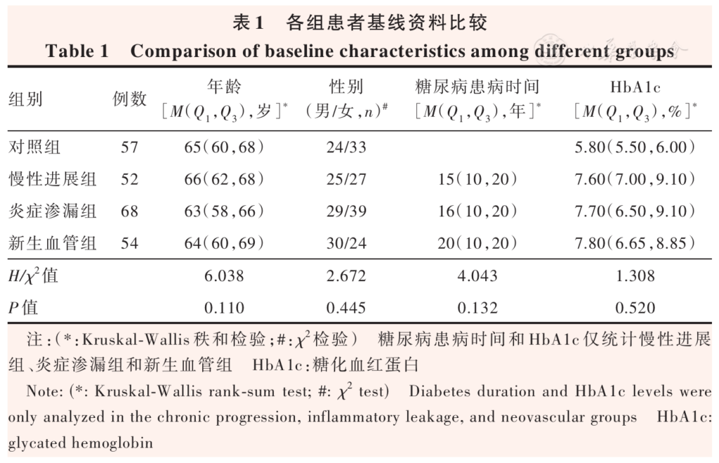
Table 1 Comparison of baseline characteristics among different groups Note: (*: Kruskal-Wallis rank-sum test; #: χ2 test) Diabetes duration and HbA1c levels were only analyzed in the chronic progression, inflammatory leakage, and neovascular groups HbA1c: glycated hemoglobin
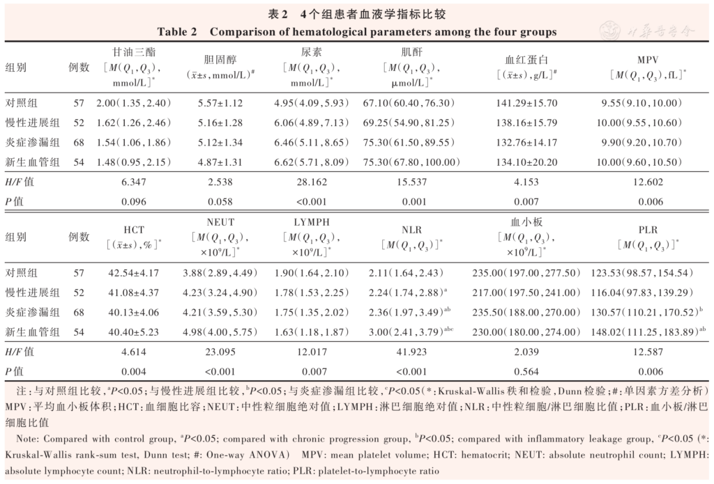
Table 2 Comparison of hematological parameters among the four groups Note: Compared with control group, aP<0.05; compared with chronic progression group, bP<0.05; compared with inflammatory leakage group, cP<0.05 (*: Kruskal-Wallis rank-sum test, Dunn test; #: One-way ANOVA) MPV: mean platelet volume; HCT: hematocrit; NEUT: absolute neutrophil count; LYMPH: absolute lymphocyte count; NLR: neutrophil-to-lymphocyte ratio; PLR: platelet-to-lymphocyte ratio
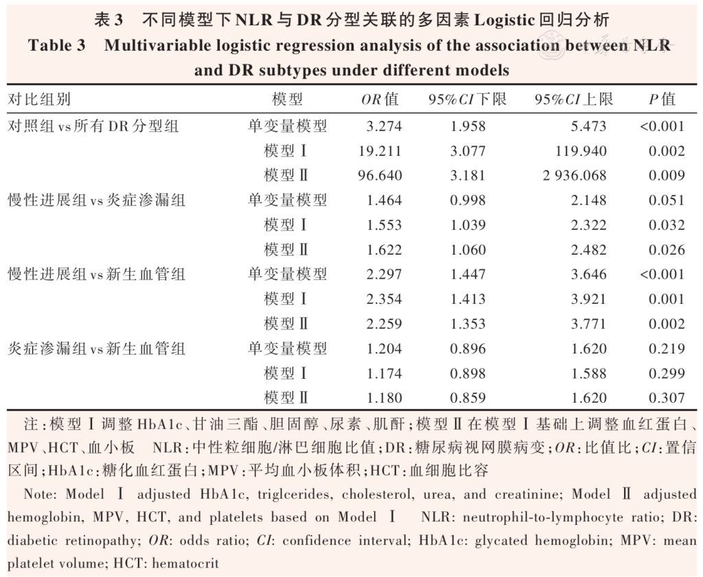
Table 3 Multivariable logistic regression analysis of the association between NLR and DR subtypes under different models Note: Model Ⅰ adjusted HbA1c, triglcerides, cholesterol, urea, and creatinine; Model Ⅱ adjusted hemoglobin, MPV, HCT, and platelets based on Model Ⅰ NLR: neutrophil-to-lymphocyte ratio; DR: diabetic retinopathy; OR: odds ratio; CI: confidence interval; HbA1c: glycated hemoglobin; MPV: mean platelet volume; HCT: hematocrit
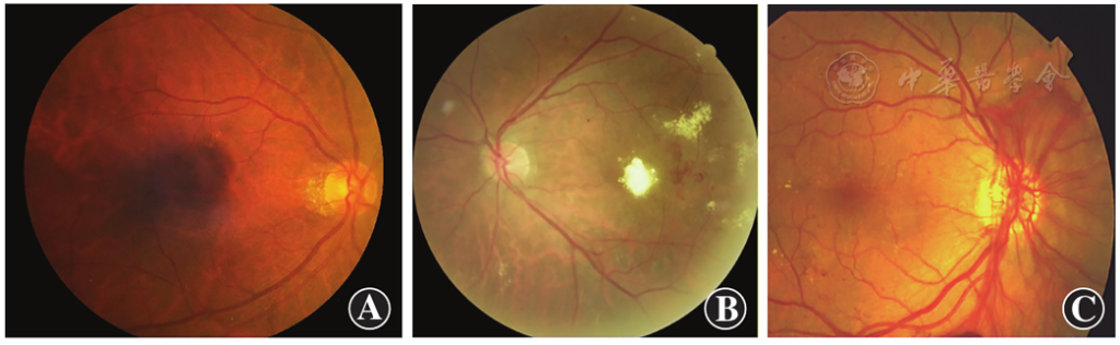
Figure 1 Schematic diagram of fundus changes in patients with different subtypes of DR A: Chronic progression type B: Inflammatory leakage type C: Neovascular type DR: diabetic retinopathy
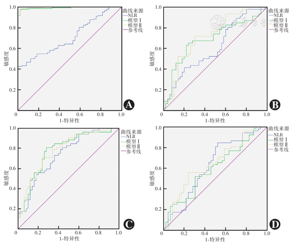
Figure 2 ROC curve analysis results for the diagnostic value of NLR in differentiating DR subtypes under different models A: Control group vs all DR subtypes combined B: Chronic progression group vs inflammatory leakage group C: Chronic progression group vs neovascular group D: Inflammatory leakage group vs neovascular group NLR: neutrophil-to-lymphocyte ratio; DR: diabetic retinopathy; ROC: receiver operating characteristic
References click to collapse
[1] Youngblood H, Robinson R, Sharma A, et al. Proteomic biomarkers of retinal inflammation in diabetic retinopathy[J]. Int J Mol Sci, 2019, 20(19):4755. DOI: 10.3390/ijms20194755.
[2] 中国微循环学会糖尿病与微循环专业委员会,中华医学会内分泌学分会基层内分泌代谢病学组,孙子林.基层糖尿病微血管疾病筛查与防治专家共识(2024)[J]. 中国全科医学, 2024, 27(32):3969-3986. DOI: 10.12114/j.issn.1007-9572.2024.0163. Microcirculation DaMPCoCSo, Endocrinology PEaMDGotCSo, Sun ZL. Expert consensus on screening and prevention of diabetic microvascular disease for primary care(2024)[J]. Chinese General Practice, 2024, 27(32):3969-3986. DOI: 10.12114/j.issn.1007-9572.2024.0163.
[3] Gardner TW, Sundstrom JM. A proposal for early and personalized treatment of diabetic retinopathy based on clinical pathophysiology and molecular phenotyping[J]. Vision Res, 2017, 139:153-160. DOI: 10.1016/j.visres.2017.03.006.
[4] Sun XJ, Zhang GH, Guo CM, et al. Associations between psycho behavioral risk factors and diabetic retinopathy: NHANES (2005—2018)[J]. Front Public Health, 2022, 10:966714. DOI: 10.3389/fpubh.2022.966714.
[5] Lin KY, Hsih WH, Lin YB, et al. Update in the epidemiology, risk factors, screening, and treatment of diabetic retinopathy[J]. J Diabetes Investig, 2021, 12(8):1322-1325. DOI: 10.1111/jdi.13480.
[6] Lobo CL, Bernardes RC, Figueira JP, et al. Three-year follow-up study of blood-retinal barrier and retinal thickness alterations in patients with type 2 diabetes mellitus and mild nonproliferative diabetic retinopathy[J]. Arch Ophthalmol, 2004, 122(2):211-217. DOI: 10.1001/archopht.122.2.211.
[7] Nunes S, Ribeiro L, Lobo C, et al. Three different phenotypes of mild nonproliferative diabetic retinopathy with different risks for development of clinically significant macular edema[J]. Invest Ophthalmol Vis Sci, 2013, 54(7):4595-4604. DOI: 10.1167/iovs.13-11895.
[8] Ribeiro L, Bandello F, Tejerina AN, et al. Characterization of retinal disease progression in a 1-year longitudinal study of eyes with mild nonproliferative retinopathy in diabetes type 2[J]. Invest Ophthalmol Vis Sci, 2015, 56(9):5698-5705. DOI: 10.1167/iovs.15-16708.
[9] Ribeiro L, Pappuru R, Lobo C, et al. Different phenotypes of mild nonproliferative diabetic retinopathy with different risks for development of macular edema (C-TRACER study)[J]. Ophthalmic Res, 2018, 59(2):59-67. DOI: 10.1159/000484666.
[10] Marques IP, Madeira MH, Messias AL, et al. Retinopathy phenotypes in type 2 diabetes with different risks for macular edema and proliferative retinopathy[J]. J Clin Med, 2020, 9(5):1433. DOI: 10.3390/jcm9051433.
[11] Cunha-Vaz J, Bernardes R. Nonproliferative retinopathy in diabetes type 2. Initial stages and characterization of phenotypes[J]. Prog Retin Eye Res, 2005, 24(3):355-377. DOI: 10.1016/j.preteyeres.2004.07.004.
[12] Xiao H, Xin W, Sun LM, et al. Comprehensive proteomic profiling of aqueous humor proteins in proliferative diabetic retinopathy[J]. Transl Vis Sci Technol, 2021, 10(6):3. DOI: 10.1167/tvst.10.6.3.
[13] Klaassen I, Van Noorden CJ, Schlingemann RO. Molecular basis of the inner blood-retinal barrier and its breakdown in diabetic macular edema and other pathological conditions[J]. Prog Retin Eye Res, 2013, 34:19-48. DOI: 10.1016/j.preteyeres.2013.02.001.
[14] Korniluk A, Koper-Lenkiewicz OM, Kamińska J, et al. Mean platelet volume (MPV):new perspectives for an old marker in the course and prognosis of inflammatory conditions[J]. Mediators Inflamm, 2019, 2019: 9213074. DOI: 10.1155/2019/9213074.
[15] Buonacera A, Stancanelli B, Colaci M, et al. Neutrophil to lymphocyte ratio: an emerging marker of the relationships between the immune system and diseases[J]. Int J Mol Sci, 2022, 23(7):3636. DOI: 10.3390/ijms23073636.
[16] Zhou Z, Chen H, Ju H, et al. Platelet indices in patients with chronic inflammatory arthritis: a systematic review and meta-analysis[J]. Platelets, 2020, 31(7):834-844. DOI: 10.1080/09537104.2019.1704714.
[17] Gasparyan AY, Ayvazyan L, Mukanova U, et al. The platelet-to-lymphocyte ratio as an inflammatory marker in rheumatic diseases[J]. Ann Lab Med, 2019, 39(4):345-357. DOI: 10.3343/alm.2019.39.4.345.
[18] Mertoglu C, Gunay M. Neutrophil-lymphocyte ratio and platelet-lymphocyte ratio as useful predictive markers of prediabetes and diabetes mellitus[J]. Diabetes Metab Syndr, 2017, 11 Suppl 1: S127-S131. DOI: 10.1016/j.dsx.2016.12.021.
[19] Serban D, Papanas N, Dascalu AM, et al. Significance of neutrophil to lymphocyte ratio (NLR) and platelet lymphocyte ratio (PLR) in diabetic foot ulcer and potential new therapeutic targets[J]. Int J Low Extrem Wounds, 2024, 23(2):205-216. DOI: 10.1177/15347346211057742.
[20] Dascalu AM, Georgescu A, Costea AC, et al. Association between neutrophil-to-lymphocyte ratio (NLR) and platelet-to-lymphocyte ratio (PLR) with diabetic retinopathy in type 2 diabetic patients[J/OL]. Cureus, 2023, 15(11):e48581[2026-03-10]. http://www.ncbi.nlm.nih.gov/pubmed/38090430. DOI: 10.7759/cureus.48581.
[21] Chua J, Lim C, Wong TY, et al. Diabetic retinopathy in the asia-pacific[J]. Asia Pac J Ophthalmol (Phila), 2018, 7(1):3-16. DOI: 10.22608/APO.2017511.
[22] Usui-Ouchi A, Kishishita S, Sakanishi Y, et al. Longitudinal renal function changes during real-world anti-vascular endothelial growth factor therapy for diabetic macular edema in Japan[J]. Jpn J Ophthalmol, 2025, 69(2):245-252. DOI: 10.1007/s10384-025-01170-x.
[23] Wang J, Xin X, Luo W, et al. Anemia and diabetic kidney disease had joint effect on diabetic retinopathy among patients with type 2 diabetes[J]. Invest Ophthalmol Vis Sci, 2020, 61(14): 25. DOI: 10.1167/iovs.61.14.25.
[24] Sato K, Jujo T, Sekine R, et al. Correlation between the outcome of vitrectomy for proliferative diabetic retinopathy and erythrocyte hematocrit level and platelet function[J]. J Clin Med, 2022, 11(17): 5055. DOI: 10.3390/jcm11175055.
[25] Grading diabetic retinopathy from stereoscopic color fundus photographs–an extension of the modified Airlie House classification. ETDRS report number 10. Early Treatment Diabetic Retinopathy Study Research Group[J]. Ophthalmology, 1991, 98(5 Suppl):786-806.
[26] Serra AM, Waddell J, Manivannan A, et al. CD11b+ bone marrow derived monocytes are the major leukocyte subset responsible for retinal capillary leukostasis in experimental diabetes in mouse and express high levels of CCR5 in the circulation[J]. Am J Pathol, 2012, 181(2):719-727. DOI: 10.1016/j.ajpath.2012.04.009.
[27] Storti F, Pulley J, Kuner P, et al. Circulating biomarkers of inflammation and endothelial activation in diabetic retinopathy[J]. Transl Vis Sci Technol, 2021, 10(12):8. DOI: 10.1167/tvst.10.12.8.
[28] Zou G, Que L, Liu Y, et al. Interplay of endothelial-mesenchymal transition, inflammation, and autophagy in proliferative diabetic retinopathy pathogenesis[J/OL]. Heliyon, 2024, 10(3):e25166[2026-03-12]. http://www.ncbi.nlm.nih.gov/pubmed/38327444. DOI: 10.1016/j.heliyon.2024.e25166.
[29] Shi Q, Wang Q, Wang Z, et al. Systemic inflammatory regulators and proliferative diabetic retinopathy: a bidirectional Mendelian randomization study[J]. Front Immunol, 2023, 14:1088778. DOI: 10.3389/fimmu.2023.1088778.
[30] Zahorec R. Neutrophil-to-lymphocyte ratio, past, present and future perspectives[J]. Bratisl Lek Listy, 2021, 122(7):474-488. DOI: 10.4149/BLL_2021_078.
[31] Duman TT, Aktas G, Atak BM, et al. Neutrophil to lymphocyte ratio as an indicative of diabetic control level in type 2 diabetes mellitus[J]. Afr Health Sci, 2019, 19(1):1602-1606. DOI: 10.4314/ahs.v19i1.35.
[32] Bulu A, Keser S. The relationship between pan-immune inflammation value and different stages of diabetic retinopathy in patients with type 2 diabetes mellitus: a prospective cross-sectional study[J]. BMC Endocr Disord, 2025, 25(1):184. DOI: 10.1186/s12902-025-02007-x.
[33] Bai Y, Huang L, Li Y, et al. Neutrophil/lymphocyte ratio and all-cause mortality in diabetic kidney disease: a retrospective cohort study[J]. J Inflamm Res, 2024, 17:10739-10753. DOI: 10.2147/JIR.S483637.
[34] Tang A, Zhang Y, Wu L, et al. Klotho’s impact on diabetic nephropathy and its emerging connection to diabetic retinopathy[J]. Front Endocrinol (Lausanne), 2023, 14:1180169. DOI: 10.3389/fendo.2023.1180169.
[35] Ebrahim H, Fiseha T, Ebrahim Y, et al. Comparison of hematological parameters between type 2 diabetes mellitus patients and healthy controls at Dessie comprehensive specialized hospital, Northeast Ethiopia: comparative cross-sectional study[J/OL]. PLoS One, 2022, 17(7):e0272145[2026-03-15]. http://www.ncbi.nlm.nih.gov/pubmed/35895700. DOI: 10.1371/journal.pone.0272145.
[36] Lu Z, Ma G, Chen L. Sitagliptin on carotid intima-media thickness in type 2 diabetes mellitus patients and anemia: a subgroup analysis of the PROLOGUE study[J]. Mediators Inflamm, 2020, 2020:8143835. DOI: 10.1155/2020/8143835.