Authors:Zhou Yanrong, Zhao Wenxin, Zhang Ming, Dang Yufei, Li Li
Corresponding author: Li Li, Email:eyelili@126.com
Published:2026-10-10
DOI: 10.3760/cma.j.cn115989-20260310-00111
ABSTRACT
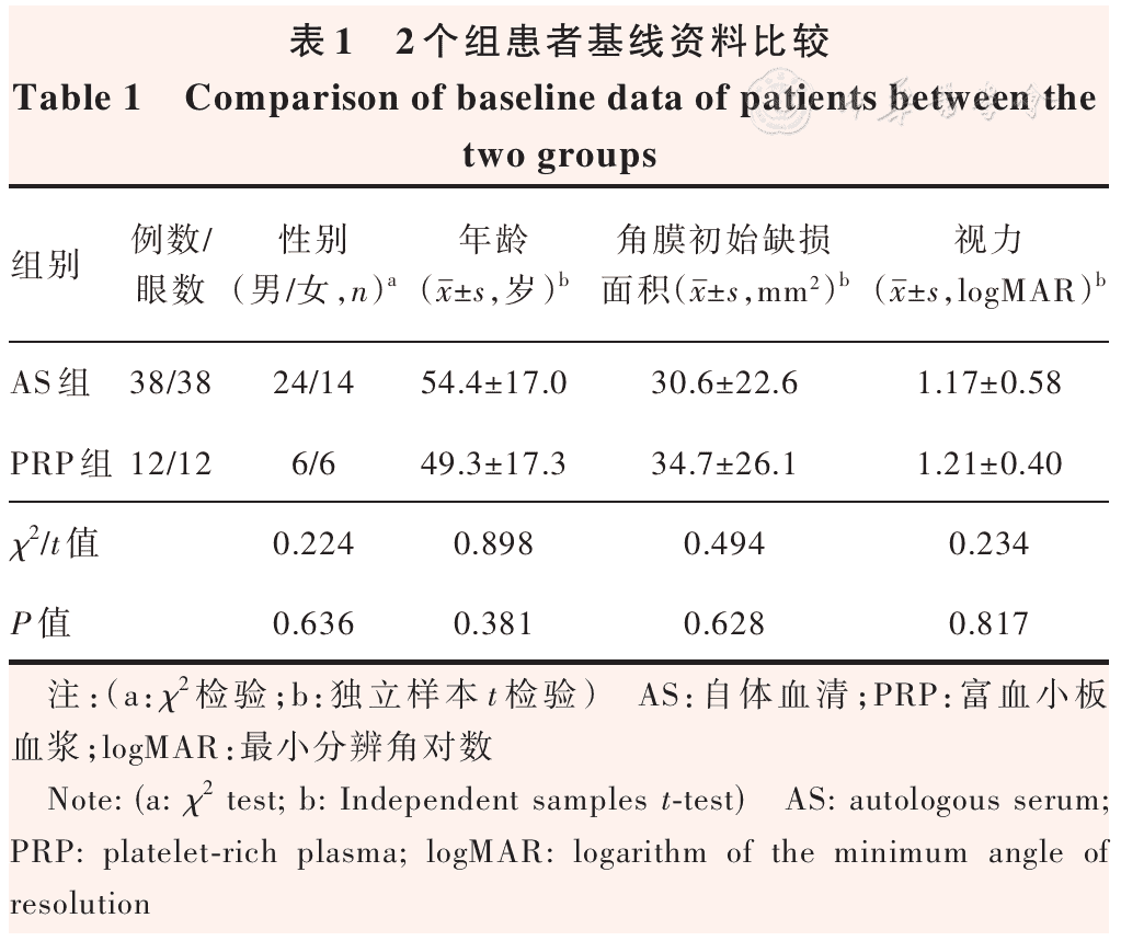
Objective To compare the efficacy of platelet-rich plasma (PRP) versus 20% autologous serum (AS) in treating refractory corneal epithelial defects (RCED).
Methods A cohort study was conducted. A total of 50 consecutive patients (50 eyes) with RCED who were treated at the First Affiliated Hospital of Xi’an Jiaotong University from December 2022 to May 2025 were enrolled. Patients were assigned to the PRP group (12 patients, 12 eyes) or the AS group (38 patients, 38 eyes) according to their treatment. Corneal epithelial defect area was quantified using ImageJ software at baseline and at 4 and 8 weeks after treatment. Visual acuity was measured at baseline and 8 weeks after treatment. Corneal epithelial healing and visual acuity improvement were compared between the two groups. This study complied with the Declaration of Helsinki, and the study protocol was approved by the Ethics Review Committee of the First Affiliated Hospital of Xi’an Jiaotong University (No. XJTU1AF2025SY-376), and all patients provided written informed consent.
Results At 4 weeks after treatment, the mean corneal epithelial healing rate in the PRP group was (0.90±0.76)mm2/day, significantly faster than (0.51±0.24)mm2/day in the AS group (t=2.69,P<0.01). The healed area in the PRP group was greater than that in the AS group at both 4 and 8 weeks after treatment (both P<0.05). At 8 weeks after treatment, the visual acuity improvement in the PRP group and AS group was 0.05(-0.18, 0.33) and 0.00(-0.10, 0.49), respectively, with no statistically significant difference (U=216.50, P=0.802). The distribution of visual acuity improvement grades also showed no significant difference between the two groups (Z=0.18, P=0.864). After propensity score matching (8 matched pairs, all standardized mean differences <0.20), the healed corneal epithelial area in the PRP group at 4 weeks was (22.79±15.37)mm2, larger than (13.64±6.31)mm2 in the AS group (t=2.31,P<0.01).
Conclusions Both PRP and AS promote corneal epithelial healing. PRP exhibits a marked therapeutic advantage over AS and may be considered a preferential option for the management of RCED.
KEYWORDS:
Platelet-rich plasma;Autologous serum;Refractory corneal epithelial defect
COPYRIGHTS:
Copyright by Chinese Medical Association
No content published by the journals of Chinese Medical Association may be reproduced or abridged without authorization. Please do not use or copy the layout and design of the journals without permission.
All articles published represent the opinions of the authors, and do not reflect the official policy of the Chinese Medical Association or the Editorial Board, unless this is clearly specified.
Authors Info & Affiliations
Zhou Yanrong
Department of Ophthalmology, The First Affiliated Hospital of Xi’an Jiaotong University, Xi’an 710000, China
Zhao Wenxin
Department of Ophthalmology, The First Affiliated Hospital of Xi’an Jiaotong University, Xi’an 710000, China
Zhang Ming
Department of Ophthalmology, The First Affiliated Hospital of Xi’an Jiaotong University, Xi’an 710000, China
Dang Yufei
Department of Ophthalmology, The First Affiliated Hospital of Xi’an Jiaotong University, Xi’an 710000, China
Li Li
Department of Ophthalmology, The First Affiliated Hospital of Xi’an Jiaotong University, Xi’an 710000, China
Figures & Tables
▼click to see the rest
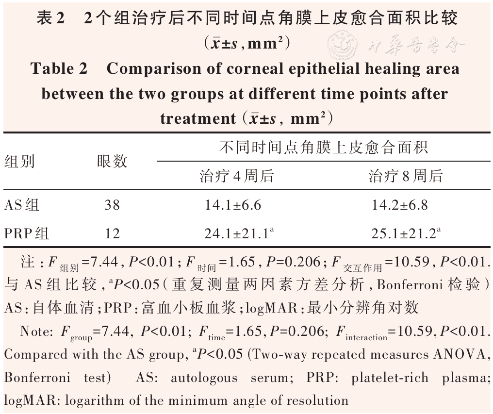
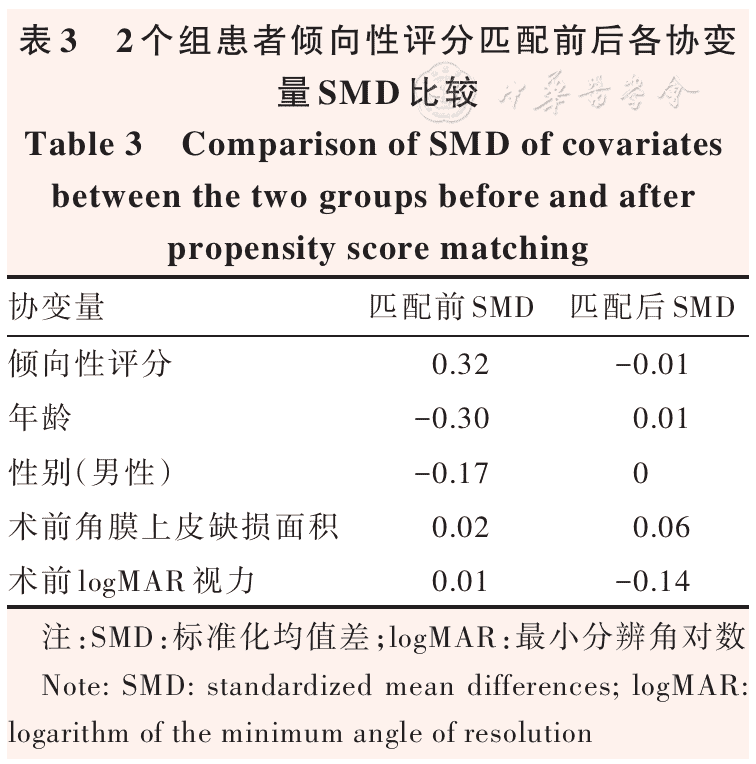
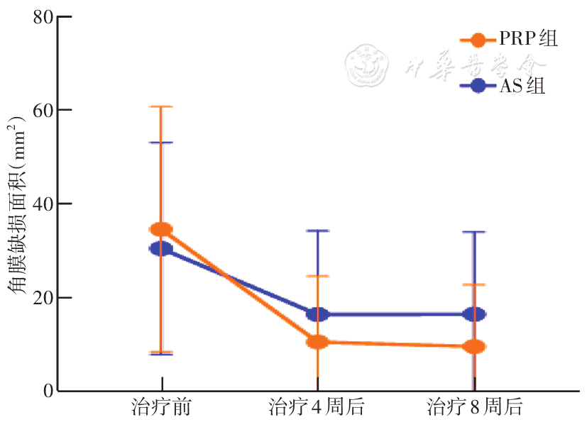
Figure 1 Comparison of corneal epithelial defect area between PRP and AS groups before and after treatment PRP: platelet-rich plasma; AS: autologous serum
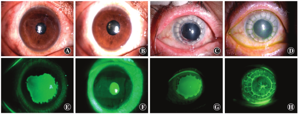
Figure 2 Corneal images before and after treatment in two groups A: Before AS treatment B: After AS treatment C: Corneal fluorescein staining before AS treatment D: Corneal fluorescein staining after AS treatment E: Before PRP treatment F: After PRP treatment G: Corneal fluorescein staining before PRP treatment H: Corneal fluorescein staining after PRP treatment AS: autologous serum; PRP: platelet-rich plasma
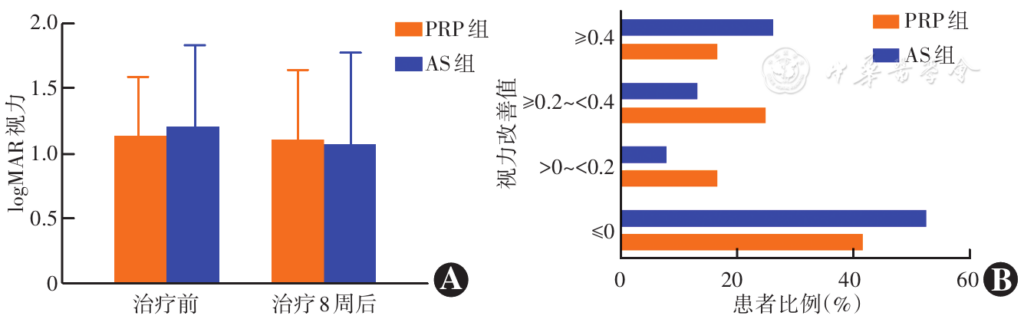
Figure 3 Comparison of visual improvement between PRP group and AS group at 8 weeks after treatment A: Comparison of visual acuity changes between the two groups before and after treatment B: Proportion of patients with different levels of visual acuity improvement in the two groups PRP: platelet-rich plasma; AS: autologous serum; logMAR: logarithm of the minimum angle of resolution
References
[1] Diaz-Valle D, Burgos-Blasco B, Gegundez-Fernandez JA, et al. Topical insulin for refractory persistent corneal epithelial defects[J]. Eur J Ophthalmol, 2021, 31(5):2280-2286. DOI: 10.1177/1120672120958307.
▼click to see more references
[2] Wilson SE, Goshe JM. Prevention and treatment of persistent epithelial defects after common refractive surgery procedures[J]. J Refract Surg, 2024, 40(2):e117-e124. DOI: 10.3928/1081597X-20240102-01.
[3] Ronci C, Ferraro AS, Lanti A, et al. Platelet-rich plasma as treatment for persistent ocular epithelial defects[J]. Transfus Apher Sci, 2015, 52(3):300-304. DOI: 10.1016/j.transci.2014.12.027.
[4] Soni NG, Jeng BH. Blood-derived topical therapy for ocular surface diseases[J]. Br J Ophthalmol, 2016, 100(1):22-27. DOI: 10.1136/bjophthalmol-2015-306842.
[5] Peng WY, He LW, Zeng P, et al. Tenonplasty combined with amniotic membrane transplantation for patients with severe ocular burns induced anterior segment necrosis[J]. J Burn Care Res, 2020, 41(3):668-673. DOI: 10.1093/jbcr/iraa016.[6] Anitua E, de la Sen-Corcuera B, Orive G, et al. Progress in the use of plasma rich in growth factors in ophthalmology: from ocular surface to ocular fundus[J]. Expert Opin Biol Ther, 2022, 22(1):31-45. DOI: 10.1080/14712598.2021.1945030.
[7] Hwang J, Chung SH, Jeon S, et al. Comparison of clinical efficacies of autologous serum eye drops in patients with primary and secondary Sjögren syndrome[J]. Cornea, 2014, 33(7):663-667. DOI: 10.1097/ICO.0000000000000147.
[8] Mahelkova G, Jirsova K, Seidler Stangova P, et al. Using corneal confocal microscopy to track changes in the corneal layers of dry eye patients after autologous serum treatment[J]. Clin Exp Optom, 2017, 100(3):243-249. DOI: 10.1111/cxo.12455.
[9] Roldan AM, De Arrigunaga S, Ciolino JB. Effect of autologous serum eye drops on corneal haze after corneal cross-linking[J]. Optom Vis Sci, 2022, 99(2):95-100. DOI: 10.1097/OPX.0000000000001839.
[10] Celebi AR, Ulusoy C, Mirza GE. The efficacy of autologous serum eye drops for severe dry eye syndrome: a randomized double-blind crossover study[J]. Graefes Arch Clin Exp Ophthalmol, 2014, 252(4):619-626. DOI: 10.1007/s00417-014-2599-1.
[11] 王文莹, 冯珺, 邓世靖,等.自体血清点眼对神经营养性角膜病变持续角膜上皮缺损的疗效评估[J].中华实验眼科杂志,2024,42(2):143-151. DOI: 10.3760/cma.j.cn115989-20221213-00584. Wang WY, Feng J, Deng SJ, et al. Evaluation of the therapeutic effect of autologous serum on neurotrophic keratopathy with persistent corneal epithelial defect[J]. Chin J Exp Ophthalmol, 2024, 42(2):143-151. DOI: 10.3760/cma.j.cn115989-20221213-00584.
[12] Arain MA, Dar AJ, Adeeb L. Autologous serum eye drops for the treatment of persistent corneal epithelial defects[J]. J Coll Physicians Surg Pak, 2013, 23(10):737-739. DOI: 10.2013/JCPSP.737739.
[13] Wróbel-Dudzińska D, Przekora A, Kazimierczak P, et al. The comparison between the composition of 100% autologous serum and 100% platelet-rich plasma eye drops and their impact on the treatment effectiveness of dry eye disease in primary Sjogren syndrome[J]. J Clin Med, 2023, 12(9):3126. DOI: 10.3390/jcm12093126.
[14] García-Conca V, Abad-Collado M, Hueso-Abancens JR, et al. Efficacy and safety of treatment of hyposecretory dry eye with platelet-rich plasma[J]. Acta Ophthalmol, 2019, 97(2):e170-e178. DOI: 10.1111/aos.13907.
[15] Alio JL, Abad M, Artola A, et al. Use of autologous platelet-rich plasma in the treatment of dormant corneal ulcers[J]. Ophthalmology, 2007, 114(7):1286-1293. DOI: 10.1016/j.ophtha.2006.10.044.
[16] Kim KM, Shin YT, Kim HK. Effect of autologous platelet-rich plasma on persistent corneal epithelial defect after infectious keratitis[J]. Jpn J Ophthalmol, 2012, 56(6):544-550. DOI: 10.1007/s10384-012-0175-y.
[17] Shtein RM, Shen JF, Kuo AN, et al. Autologous serum-based eye drops for treatment of ocular surface disease: a report by the American Academy of Ophthalmology[J]. Ophthalmology, 2020, 127(1):128-133. DOI: 10.1016/j.ophtha.2019.08.018.
[18] Riestra AC, Alonso H, Merayo L. Platelet rich plasma in ocular surface[J]. Archivos de la Sociedad Española de Oftalmología (English Edition), 2016, 91(10):475-490. DOI: 10.1016/j.oftale.2016.06.003.
[19] Özlük İ, Yüksel B, Küsbeci T. Comparison of autologous serum and platelet-rich plasma in the treatment of severe dry eye and persistent epithelial defects[J]. Cont Lens Anterior Eye, 2024, 47(6):102247. DOI: 10.1016/j.clae.2024.102247.
[20] Medeiros CS, Marino GK, Santhiago MR, et al. The corneal basement membranes and stromal fibrosis[J]. Invest Ophthalmol Vis Sci, 2018, 59(10):4044-4053. DOI: 10.1167/iovs.18-24428.
[21] Kwon JE, Kang C, Moghtader A, et al. Emerging treatments for persistent corneal epithelial defects[J]. Vision (Basel), 2025, 9(2):26. DOI: 10.3390/vision9020026.
[22] Sanchez-Avila RM, Merayo-Lloves J, Riestra AC, et al. Plasma rich in growth factors membrane as coadjuvant treatment in the surgery of ocular surface disorders[J]. Medicine (Baltimore), 2018, 97(17):e0242. DOI: 10.1097/MD.0000000000010242.
[23] Anitua E, de la Fuente M, Muruzabal F, et al. Plasma rich in growth factors (PRGF) eye drops stimulates scarless regeneration compared to autologous serum in the ocular surface stromal fibroblasts[J]. Exp Eye Res, 2015, 135:118-126. DOI: 10.1016/j.exer.2015.02.016.